
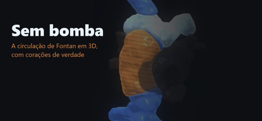
Fontan em 3D: o caminho até o pulmão que não passa pelo coração
Na circulação de Fontan com conexão cavopulmonar total, o sangue das veias cavas vai direto para a artéria pulmonar, sem ventrículo nenhum para empurrá-lo, e por isso a pressão dentro do tórax pesa tanto no pós-operatório. Usei a ressonância de uma criança de 6 anos para mostrar o caminho: os ventrículos saem de cena, o exame é desenrolado ao longo do sangue e a régua marca 40 mm de bomba no coração normal contra zero no Fontan. Na revisão, uma medida minha encolheu o tubo em três milímetros. Estava errada, e conto como percebi.
📅 Publicado em 1º de novembro de 2026
Navegue pelo Artigo
Do plantão ao exame desenrolado, e do exame à conduta na UTI.
- 1. Por que isso importa para quem trabalha na ponta
- 2. Assista: o caminho sem bomba
- 3. Explore os corações em 3D
- 4. O corte que mostra o tubo
- 5. De onde vem o material
- 6. Como o filme foi construído
- 7. Os números, sem filtro
- 8. A armadilha: o tubo que eu quase medi errado
- 9. Na UTI: proteger o gargalo
- 10. O que fazer na segunda-feira
- 11. Considerações Finais
- 12. Referências
1. Por que isso importa para quem trabalha na ponta
Pós-operatório imediato de Fontan, primeira madrugada na UTI. A pressão venosa central está alta, a saturação caiu um pouco, o dreno pleural não para de drenar e alguém sugere aumentar a PEEP “para recrutar”. Se a imagem mental da circulação de Fontan for a de um coração com um ventrículo a menos, a PEEP parece inofensiva. Só que o sangue sobe da veia cava para o pulmão sem bomba nenhuma no caminho, e toda pressão a mais dentro do tórax empurra contra esse fluxo.
Desde a cirurgia descrita por Fontan e Baudet em 1971 [2] e da conexão cavopulmonar total proposta por de Leval em 1988 [3], a frase dos livros é a mesma: no Fontan, o sangue chega ao pulmão sem passar por um ventrículo. No quadro, o desenho costuma virar um emaranhado de setas azuis (já desenhei muitos), e a frase continua abstrata.
Este é o segundo filme da série Cinematic Education feito com corações reais. No anterior, o da transposição das grandes artérias, a criança já tinha a cirurgia de Glenn, em que a veia cava superior vai direto para a artéria pulmonar. Aqui está o passo seguinte: o Fontan, em que a veia cava inferior também vai.
2. Assista: o caminho sem bomba
Ligue o som. Duas vozes conduzem o filme por 1 minuto e 28 segundos, e a imagem acompanha cada frase. A versão em inglês, com as mesmas vozes, está na aba EN.
Coração normal (4 anos) × circulação de Fontan após Glenn (6 anos). Dados: HVSMR-2.0 (Pace et al., Sci Data 2024) · CC BY 4.0 · modificado. Trilha: “Convergence” — Scott Buckley · CC BY 4.0.
O que você vai ver
O tubo atrás do coração. O filme abre por trás. A veia cava inferior acende de baixo para cima: em azul, a parte nativa; em cobre com anéis, o conduto do Fontan, que segue até a artéria pulmonar.
Os ventrículos se desprendem. Na frase “sem passar por nenhum ventrículo”, os ventrículos e a aorta saem do coração e somem. O caminho até o pulmão continua inteiro. Nesta ressonância, a artéria pulmonar não toca nenhum ventrículo: zero voxels de contato.
Do Glenn ao Fontan. O coração do episódio da transposição aparece ao lado, para comparar o estágio anterior (só a cava superior chega à pulmonar) com este (as duas cavas chegam).
O disco e o corte. Um disco de corte desliza pela veia cava, perpendicular ao caminho do sangue. Ao lado, aparece a própria ressonância naquele plano: irregular na veia nativa, redonda no conduto, com 22 mm (seção 4).
O caminho desenrolado. A linha central do sangue sai do coração e se estica numa coluna, com a ressonância real dentro. No normal, 40 mm do caminho passam pelo ventrículo direito; no Fontan, zero.
O gargalo e a fenestração. Um esquema, rotulado como esquema, mostra a pressão venosa e a respiração empurrando o sangue, o leito pulmonar como gargalo e a fenestração aliviando a pressão à custa de saturação.
3. Explore os corações em 3D
Os mesmos dois corações do filme estão aqui para você girar, ampliar e tocar. Comece pelo botão Caminho ao pulmão. No Fontan, ficam só as veias cavas, o conduto e a artéria pulmonar, num caminho contínuo, sem ventrículo. No coração normal, o mesmo botão deixa o caminho interrompido, porque ali o sangue precisa passar pelo átrio e pelo ventrículo direitos. É nesse buraco que, no coração normal, fica a bomba.
Agora, o caminho é seu.
Gire os dois corações juntos, amplie e toque em uma estrutura para identificá-la. Use “Caminho ao pulmão” para ver só as veias, o conduto e a artéria pulmonar: no Fontan, nenhum ventrículo fica no caminho. Os dois modelos usam a mesma escala em milímetros.
O modelo só é baixado quando você abre.
4. O corte que mostra o tubo
O conjunto de dados não separa o conduto da veia: para os especialistas que fizeram a segmentação, tudo o que vai do fígado à artéria pulmonar é “veia cava inferior”. Na primeira versão do filme, a coluna aparecia inteira em uma cor só, e a pergunta da revisão clínica foi direta: onde está o tubo?
A resposta veio do próprio exame. Ao longo do caminho do sangue, a cada meio milímetro, o programa corta a ressonância num plano perpendicular ao fluxo e mede a parede em 48 direções. A veia nativa tem seção irregular, com mais de um lúmen e diâmetro médio de 18,5 mm. Num trecho de 36,5 mm, a seção fica redonda, com parede contínua e calibre constante de 22,5 mm em média. Esse é o conduto, e é assim que ele aparece no filme e no modelo acima, em cobre.

Ressonância original reamostrada no plano perpendicular à linha central do lúmen, janela de 44 mm. Contornos: azul, veia nativa; cobre, conduto; cinza, átrio. HVSMR-2.0 [1] · CC BY 4.0 · modificado.
💡 Por que “inferido” e não “medido no registro”
Os metadados do caso dizem apenas “Glenn + Fontan”. Tubo redondo, parede contínua, calibre constante e posição por fora do átrio, separado dele por cerca de 1 mm, formam o padrão de um conduto extracardíaco. Num túnel lateral, o canal correria por dentro do átrio. Ainda assim, é leitura de imagem; o registro não diz isso, e por esse motivo o filme e o modelo trazem “inferido da RM”. Pelo mesmo exame, não se vê fenestração aberta: com voxel de 0,7 mm, um orifício típico de 4 mm ocuparia uns seis voxels. Uma fenestração muito pequena, ou já fechada, não pode ser excluída.
5. De onde vem o material
Os corações vêm do HVSMR-2.0, um conjunto público de 60 ressonâncias cardíacas de pacientes com cardiopatia congênita, publicado em 2024 na Scientific Data por pesquisadores do MIT, do Massachusetts General Hospital e do Boston Children’s Hospital [1]. Cada exame traz as câmaras e os grandes vasos segmentados manualmente por especialistas, sob licença CC BY 4.0 (uso educacional e comercial permitido com crédito e indicação das modificações).
Para escolher o caso, o programa mediu as conexões reais de todos os candidatos com Fontan. Em uma criança de 6 anos, as duas veias cavas chegam à artéria pulmonar (245 e 476 voxels de contato), a cava inferior quase não toca o átrio (9 voxels) e a artéria pulmonar não toca nenhum ventrículo. É uma conexão cavopulmonar total limpa, e a criança tem idade próxima à do coração normal de comparação (4 anos) e à do caso de Glenn do episódio anterior (2 anos). Os exames são desidentificados.
6. Como o filme foi construído
Cada quadro foi calculado a partir dos dados, num motor 3D próprio escrito em Python para a placa de vídeo (ModernGL), com roteiro e decisões guiados pela Metodologia AIMED. Nenhum quadro saiu de gerador de vídeo.
Os ventrículos que se desprendem
Os ventrículos saem do coração sem truque de edição: cada estrutura do exame é uma superfície separada, e o motor apenas deixa de desenhar ventrículos e aorta. O que sobra, ligado de ponta a ponta, é o que os dados mostram: veias cavas, conduto e artéria pulmonar.
O caminho desenrolado
O programa traça a linha central do lúmen da veia cava inferior até a artéria pulmonar, passando pelos pontos onde as estruturas se tocam de verdade, e reamostra a ressonância ao longo dela, num referencial que não gira à toa (transporte paralelo). É a reformatação curva, a mesma técnica que o radiologista usa para “esticar” uma coronária. Os 40 mm e o zero na tela saem dessa mesma conta.
O tubo visível
O conduto foi separado da veia pelo critério da seção 4 e ganhou material próprio: cobre, com anéis a cada 2,5 mm, como uma prótese. A veia nativa ficou em azul e a artéria pulmonar em gelo. Também passou a valer a oclusão real: o tubo, que está atrás do coração, aparece atrás dele, e o que fica escondido aparece só como transparência fraca.
Vozes, legendas e trilha
O diálogo foi escrito a partir das medidas conferidas e das fontes da seção 9, gravado com duas vozes sintéticas (Kore e Zubenelgenubi, no Google AI Studio), numa geração para português e outra para inglês. Um modelo de reconhecimento de fala (faster-whisper) devolveu o tempo de cada palavra; a legenda foi alinhada palavra por palavra ao texto escrito, e onde o reconhecedor errou (o nome da metodologia, por exemplo), o tempo foi interpolado entre as palavras vizinhas. A trilha “Convergence”, de Scott Buckley (CC BY 4.0), cresce no desenrolar e abaixa sob as vozes.
7. Os números, sem filtro
| Medida (calculada nos dados) | Normal (4 anos) | Fontan (6 anos) | Fonte | Leitura |
|---|---|---|---|---|
| Caminho do sangue da VCI ao ramo da AP | 151 mm | 108,5 mm | HVSMR-2.0 [1] | ✓ linha central |
| Trecho do caminho dentro de ventrículo | 40,5 mm (VD) | 0 mm | HVSMR-2.0 [1] | → sem bomba |
| Contato da AP com ventrículos | VD: 383 voxels | 0 voxels | HVSMR-2.0 [1] | → Fontan |
| Veia cava superior drena para | átrio direito (380 voxels) | artéria pulmonar (245) | HVSMR-2.0 [1] | → Glenn |
| Veia cava inferior drena para | átrio direito (609 voxels) | artéria pulmonar (476) · átrio (9) | HVSMR-2.0 [1] | → cavopulmonar total |
| Conduto: comprimento e diâmetro médio | — | 36,5 mm · 22,5 mm | cálculo próprio | ✓ seção perpendicular |
| Irregularidade da seção (desvio dos raios) | — | conduto 0,10 · veia nativa 0,25 | cálculo próprio | ✓ tubo redondo |
💡 Traduzindo para o chão da UTI
A segunda linha resume o episódio. No coração normal, 40 mm do caminho até o pulmão passam por dentro de uma câmara que contrai. No Fontan, nenhum milímetro passa. O que empurra o sangue pelo pulmão é a pressão nas veias, gerada lá atrás pelo ventrículo único e ajudada pela respiração, e por isso a resistência pulmonar decide o débito [4].
8. A armadilha: o tubo que eu quase medi errado
Número na tela dá autoridade ao filme, inclusive quando está errado (e aí o espectador memoriza o erro com a mesma confiança). Os três problemas desta revisão tinham uma coisa em comum: para quem não conhece a anatomia, o filme parecia certo.
⚠ Os três problemas que a revisão encontrou
1. O tubo aparecia na frente do coração. Para o caminho venoso brilhar, a primeira versão o desenhava por cima de tudo. Na vista anterior, o conduto, que fica atrás, surgia na frente do coração. Ficava bonito. Anatomicamente, estava errado. Hoje vale a oclusão real, e o que está escondido aparece só como transparência fraca.
2. O tubo que ninguém via. O conjunto de dados dá um rótulo só para a veia nativa, o conduto e a anastomose. O filme mostrava uma coluna única, e a pergunta da revisão clínica (“não achei o tubo”) apontou isso. O conduto passou a ser delimitado pela própria imagem (seção 4).
3. A medida que encolheu o tubo. Ao conferir os 22 mm, usei primeiro o diâmetro da maior esfera que cabe dentro do lúmen, e o resultado foi 18–19 mm. Por algumas horas, achei que a fala do filme estava errada. O erro era meu: a esfera inscrita mede a menor distância até a parede e é puxada para baixo por qualquer saliência da segmentação. Medindo a seção perpendicular em todas as direções, o conduto tem 22–23 mm, o mesmo que o corte axial já mostrava. Se eu tivesse confiado no número menor, o filme teria trocado um valor certo por um errado, e com a aparência de quem conferiu.
⚠ Limites deste filme
As superfícies mostram só o lúmen; o miocárdio ficou de fora. O caminho segue a linha central do lúmen, sem medida de fluxo. A respiração, o gargalo e a fenestração aparecem como esquemas rotulados, sem simulação por trás. O conduto extracardíaco e a ausência de fenestração aberta são inferências da ressonância. É um caso só, longe de cobrir o espectro dos Fontans. Serve para ensinar; para diagnóstico ou planejamento cirúrgico, não serve.
9. Na UTI: proteger o gargalo
Na circulação de Fontan não há bomba entre as veias e o pulmão; por isso o fluxo pulmonar depende de duas coisas: da pressão nas veias e da pressão dentro do tórax.
A respiração ajuda. Na conexão cavopulmonar total, o fluxo pulmonar é fásico: aumenta na inspiração espontânea e chega a inverter na manobra de Valsalva [5]. O fluxo das veias hepáticas na inspiração é 3,4 vezes o da expiração no Fontan, contra 1,7 em controles [6].
A pressão positiva atrapalha. Em crianças após Fontan, trocar a ventilação com pressão positiva por ventilação com pressão negativa aumentou o fluxo pulmonar em 42–54%, só pelo aumento do volume sistólico, e o ganho sumiu ao voltar para a pressão positiva [7]. A extubação precoce, ainda na sala, associou-se a menor pressão na artéria pulmonar e no átrio nas primeiras 12 horas, num estudo retrospectivo com seleção de pacientes [8].
A resistência pulmonar é o gargalo. Hipóxia e acidose aumentam a resistência vascular pulmonar [9], e no Fontan isso cai direto sobre o débito [4].
A fenestração é a válvula de alívio. Um pequeno orifício do conduto para o átrio deixa sair sangue venoso quando a pressão sobe: sustenta o débito e limita a pressão venosa, à custa de shunt da direita para a esquerda (saturação média de 86% na série original) [10]. Num ensaio randomizado com 49 pacientes de risco padrão, a fenestração reduziu em 55% a drenagem torácica e em 41% o tempo de internação [11]. Nesta ressonância, não se vê fenestração aberta.
Um exemplo concreto: “a PEEP que fecha o caminho”
Na madrugada do pós-operatório, a cava está alta e a saturação baixou. O reflexo de quem pensa num coração comum é subir a PEEP para recrutar. No Fontan, a pergunta certa é outra: qual é o mínimo de pressão positiva que mantém o pulmão aberto? Atelectasia e hiperinsuflação aumentam a resistência pulmonar, e qualquer pressão a mais no tórax empurra contra um fluxo que não tem bomba. O mesmo raciocínio leva a extubar cedo, quando possível, para que a inspiração volte a puxar o sangue [7, 8, 12].
⚠ Magnitude da evidência
O dado mais forte sobre ventilação (pressão negativa por couraça) vem de 18 crianças e de um modo ventilatório fora da rotina [7]. A extubação precoce vem de um estudo retrospectivo [8]. Por isso o texto e o filme falam em “mínimo de pressão positiva” e “extubar cedo” como direção, sem prescrever valores de PEEP ou de pressão média. O manejo completo está na declaração científica da AHA [12].
10. O que fazer na segunda-feira
Antes do próximo Fontan na UTI
Use IA com método — e com revisão clínica
A Metodologia AIMED ensina médicos a usar inteligência artificial para produzir material de ensino e de trabalho com rigor: fonte primária, conferência e responsabilidade de quem assina.
Considerações Finais
O episódio começou com uma pergunta de beira de leito (onde está o tubo?) e terminou com duas correções minhas: separar o conduto pela própria imagem e desistir de um número que parecia mais rigoroso do que era.
Usei IA para escrever o código, gerar as vozes e rodar os cálculos. O caso, o recorte, as fontes da conduta e a revisão ficaram comigo; os dados são de especialistas que segmentaram cada exame à mão e os deixaram abertos para outros usarem. Este é o terceiro episódio da série; o anterior, com corações reais, foi o da transposição das grandes artérias, e todos ficam reunidos na coleção Cinematic Education.
💡 Connecting the Dots: que no Fontan não há ventrículo a caminho do pulmão cai em prova de residência. A consequência de beira de leito (o sangue chega ao pulmão empurrado pela pressão das veias e puxado pela inspiração, e a pressão positiva atrapalha justamente esse trajeto) costuma ser aprendida tarde, às vezes na primeira madrugada de pós-operatório. Os 40 mm e o zero da tela tentam antecipar essa madrugada. Do lado do processo, ficou um aviso: a medida errada tinha mais cara de conferida do que a certa, e quem a derrubou foi uma pergunta clínica. O próximo episódio sai na semana que vem.
Referências
- Pace DF, Contreras HTM, Romanowicz J, et al. HVSMR-2.0: A 3D cardiovascular MR dataset for whole-heart segmentation in congenital heart disease. Sci Data. 2024;11(1):721. Disponível em: https://doi.org/10.1038/s41597-024-03469-9 · Dados (CC BY 4.0): https://doi.org/10.6084/m9.figshare.c.7074755.v2
- Fontan F, Baudet E. Surgical repair of tricuspid atresia. Thorax. 1971;26(3):240-8. PMID: 5089489. Disponível em: https://pubmed.ncbi.nlm.nih.gov/5089489/
- de Leval MR, Kilner P, Gewillig M, Bull C. Total cavopulmonary connection: a logical alternative to atriopulmonary connection for complex Fontan operations. J Thorac Cardiovasc Surg. 1988;96(5):682-95. PMID: 3184963. Disponível em: https://pubmed.ncbi.nlm.nih.gov/3184963/
- Gewillig M. The Fontan circulation. Heart. 2005;91(6):839-46. Disponível em: https://doi.org/10.1136/hrt.2004.051789
- Redington AN, Penny D, Shinebourne EA. Pulmonary blood flow after total cavopulmonary shunt. Br Heart J. 1991;65(4):213-7. Disponível em: https://doi.org/10.1136/hrt.65.4.213
- Hsia TY, Khambadkone S, Redington AN, et al. Effects of respiration and gravity on infradiaphragmatic venous flow in normal and Fontan patients. Circulation. 2000;102(19 Suppl 3):III148-53. Disponível em: https://doi.org/10.1161/01.cir.102.suppl_3.iii-148
- Shekerdemian LS, Bush A, Shore DF, et al. Cardiopulmonary interactions after Fontan operations: augmentation of cardiac output using negative pressure ventilation. Circulation. 1997;96(11):3934-42. Disponível em: https://doi.org/10.1161/01.cir.96.11.3934
- Morales DLS, Carberry KE, Heinle JS, et al. Extubation in the operating room after Fontan’s procedure: effect on practice and outcomes. Ann Thorac Surg. 2008;86(2):576-81. Disponível em: https://doi.org/10.1016/j.athoracsur.2008.02.010
- Rudolph AM, Yuan S. Response of the pulmonary vasculature to hypoxia and H+ ion concentration changes. J Clin Invest. 1966;45(3):399-411. Disponível em: https://doi.org/10.1172/JCI105355
- Bridges ND, Lock JE, Castaneda AR. Baffle fenestration with subsequent transcatheter closure. Modification of the Fontan operation for patients at increased risk. Circulation. 1990;82(5):1681-9. Disponível em: https://doi.org/10.1161/01.cir.82.5.1681
- Lemler MS, Scott WA, Leonard SR, et al. Fenestration improves clinical outcome of the Fontan procedure: a prospective, randomized study. Circulation. 2002;105(2):207-12. Disponível em: https://doi.org/10.1161/hc0202.102237
- Rychik J, Atz AM, Celermajer DS, et al. Evaluation and management of the child and adult with Fontan circulation: a scientific statement from the American Heart Association. Circulation. 2019;140(6):e234-e284. PMID: 31256636. Disponível em: https://pubmed.ncbi.nlm.nih.gov/31256636/
- Buckley S. Convergence (trilha). Licença CC BY 4.0. Disponível em: https://www.scottbuckley.com.au/library/
Fontan in 3D: the path to the lungs that bypasses the heart
In a Fontan circulation with a total cavopulmonary connection, blood from the venae cavae goes straight to the pulmonary artery, with no ventricle to push it, which is why pressure inside the chest matters so much after surgery. I used the MRI of a 6-year-old to show the path: the ventricles leave the scene, the scan is unrolled along the blood, and the ruler reads 40 mm of pump in the normal heart against zero in the Fontan. During the review, one of my measurements shrank the tube by three millimetres. It was wrong, and I explain how I noticed.
📅 Published November 1, 2026
Table of Contents
From the bedside to the unrolled scan, and from the scan to ICU management.
- 1. Why this matters at the bedside
- 2. Watch: the path with no pump
- 3. Explore the hearts in 3D
- 4. The slice that shows the tube
- 5. Where the material comes from
- 6. How the film was built
- 7. The numbers, unfiltered
- 8. The trap: the tube I almost measured wrong
- 9. In the ICU: protect the bottleneck
- 10. What to do on Monday
- 11. Final Considerations
- 12. References
1. Why this matters at the bedside
Immediate post-op Fontan, first night in the ICU. Central venous pressure is high, saturation has dropped a little, the chest drain keeps draining, and someone suggests raising the PEEP “to recruit”. If your mental image of the Fontan circulation is a heart with one ventricle missing, the PEEP looks harmless. But blood rises from the vena cava to the lungs with no pump on the way, and any extra pressure inside the chest pushes against that flow.
Since the operation described by Fontan and Baudet in 1971 [2] and the total cavopulmonary connection proposed by de Leval in 1988 [3], the textbook sentence has been the same: in a Fontan, blood reaches the lungs without passing through a ventricle. On a whiteboard, the drawing usually turns into a tangle of blue arrows (I have drawn plenty), and the sentence stays abstract.
This is the second Cinematic Education film made with real hearts. In the previous one, on transposition of the great arteries, the child already had a Glenn operation, in which the superior vena cava goes straight to the pulmonary artery. Here is the next step: the Fontan, in which the inferior vena cava goes there too.
2. Watch: the path with no pump
Turn the sound on. Two voices lead the film for 1 minute and 33 seconds, in English, and the image follows each sentence.
Normal heart (age 4) × Fontan circulation after Glenn (age 6). Data: HVSMR-2.0 (Pace et al., Sci Data 2024) · CC BY 4.0 · modified. Music: “Convergence” — Scott Buckley · CC BY 4.0.
What you will see
The tube behind the heart. The film opens from behind. The inferior vena cava lights up from the bottom: in blue, the native part; in copper with rings, the Fontan conduit, running up to the pulmonary artery.
The ventricles float away. On “without passing through any ventricle”, the ventricles and the aorta leave the heart and disappear. The path to the lungs stays intact. In this MRI, the pulmonary artery touches no ventricle: zero contact voxels.
From Glenn to Fontan. The heart from the transposition episode appears alongside, to compare the previous stage (only the superior vena cava reaches the pulmonary artery) with this one (both cavae reach it).
The disc and the slice. A cutting disc slides along the vena cava, perpendicular to the blood’s path. Beside it, the MRI itself in that plane: irregular in the native vein, round in the conduit, 22 mm wide (section 4).
The path, unrolled. The blood’s centreline leaves the heart and stretches into a column, with the real MRI inside. In the normal heart, 40 mm of the path runs through the right ventricle; in the Fontan, zero.
The bottleneck and the fenestration. A schematic, labelled as a schematic, shows venous pressure and breathing pushing the blood, the pulmonary bed as the bottleneck, and the fenestration relieving pressure at the cost of saturation.
3. Explore the hearts in 3D
The same two hearts from the film are here for you to rotate, zoom and tap. Start with the Path to the lungs button. In the Fontan, only the venae cavae, the conduit and the pulmonary artery remain, in one continuous path with no ventricle. In the normal heart, the same button leaves the path broken, because there blood has to pass through the right atrium and ventricle. That gap is where, in a normal heart, the pump sits.
Now, follow the path yourself.
Rotate both hearts together, zoom in and tap a structure to identify it. Use “Path to the lungs” to keep only the veins, the conduit and the pulmonary artery: in the Fontan, no ventricle stands in the way. Both models use the same scale in millimetres.
The model downloads only when you open it.
4. The slice that shows the tube
The dataset does not separate the conduit from the vein: for the experts who did the segmentation, everything from the liver to the pulmonary artery is “inferior vena cava”. In the first version of the film, the column appeared in a single colour, and the clinical review asked the obvious question: where is the tube?
The answer came from the scan itself. Along the blood’s path, every half millimetre, the program cuts the MRI in a plane perpendicular to the flow and measures the wall in 48 directions. The native vein has an irregular section, with more than one lumen and a mean diameter of 18.5 mm. Over a 36.5 mm stretch, the section becomes round, with a continuous wall and a constant calibre of 22.5 mm on average. That is the conduit, and that is how it appears in the film and in the model above, in copper.

Original MRI resampled in the plane perpendicular to the lumen centreline, 44 mm window. Contours: blue, native vein; copper, conduit; grey, atrium. HVSMR-2.0 [1] · CC BY 4.0 · modified.
💡 Why “inferred” and not “measured in the registry”
The case metadata only say “Glenn + Fontan”. A round tube, continuous wall, constant calibre and a position outside the atrium, separated from it by about 1 mm, make up the pattern of an extracardiac conduit. A lateral tunnel would run inside the atrium. Still, it is image reading; the registry does not say so, which is why the film and the model carry “inferred from the MRI”. The same scan shows no open fenestration: with 0.7 mm voxels, a typical 4 mm hole would span about six voxels. A very small fenestration, or one already closed, cannot be ruled out.
5. Where the material comes from
The hearts come from HVSMR-2.0, a public set of 60 cardiac MR scans of patients with congenital heart disease, published in 2024 in Scientific Data by researchers from MIT, Massachusetts General Hospital and Boston Children’s Hospital [1]. Each scan comes with the chambers and great vessels manually segmented by experts, under a CC BY 4.0 licence (educational and commercial use allowed with credit and indication of changes).
To choose the case, the program measured the real connections of every Fontan candidate. In a 6-year-old, both venae cavae reach the pulmonary artery (245 and 476 contact voxels), the inferior cava barely touches the atrium (9 voxels) and the pulmonary artery touches no ventricle. It is a clean total cavopulmonary connection, and the child is close in age to the normal comparison heart (age 4) and to the Glenn case from the previous episode (age 2). The scans are de-identified.
6. How the film was built
Every frame was computed from the data, in a custom 3D engine written in Python for the graphics card (ModernGL), with script and decisions guided by the AIMED Methodology. No frame came out of a video generator.
The ventricles that float away
The ventricles leave the heart without any editing trick: each structure in the scan is a separate surface, and the engine just stops drawing the ventricles and the aorta. What remains, connected end to end, is what the data show: venae cavae, conduit and pulmonary artery.
The path, unrolled
The program traces the lumen centreline from the inferior vena cava to the pulmonary artery, through the points where the structures actually touch, and resamples the MRI along it, in a frame that does not spin for no reason (parallel transport). This is curved planar reformation, the same technique a radiologist uses to “straighten” a coronary artery. The 40 mm and the zero on screen come from that same computation.
The visible tube
The conduit was separated from the vein using the criterion in section 4 and given its own material: copper, with rings every 2.5 mm, like a graft. The native vein is blue and the pulmonary artery ice-white. Real occlusion now applies too: the tube, which lies behind the heart, appears behind it, and what is hidden shows only as a faint transparency.
Voices, captions and music
The dialogue was written from the verified measurements and the sources in section 9, and recorded with two synthetic voices (Kore and Zubenelgenubi, in Google AI Studio), in one generation for Portuguese and another for English. A speech-recognition model (faster-whisper) returned the timing of every word; the caption was aligned word by word to the written text, and where the recognizer got a word wrong (the methodology’s name, for example), the timing was interpolated between the neighbouring words. The music, “Convergence” by Scott Buckley (CC BY 4.0), builds during the unrolling and ducks under the voices.
7. The numbers, unfiltered
| Measurement (computed from the data) | Normal (age 4) | Fontan (age 6) | Source | Reading |
|---|---|---|---|---|
| Blood path from IVC to a PA branch | 151 mm | 108.5 mm | HVSMR-2.0 [1] | ✓ centreline |
| Stretch of the path inside a ventricle | 40.5 mm (RV) | 0 mm | HVSMR-2.0 [1] | → no pump |
| PA contact with ventricles | RV: 383 voxels | 0 voxels | HVSMR-2.0 [1] | → Fontan |
| Superior vena cava drains into | right atrium (380 voxels) | pulmonary artery (245) | HVSMR-2.0 [1] | → Glenn |
| Inferior vena cava drains into | right atrium (609 voxels) | pulmonary artery (476) · atrium (9) | HVSMR-2.0 [1] | → total cavopulmonary |
| Conduit: length and mean diameter | — | 36.5 mm · 22.5 mm | own calculation | ✓ perpendicular section |
| Section irregularity (spread of radii) | — | conduit 0.10 · native vein 0.25 | own calculation | ✓ round tube |
💡 Translating to the ICU floor
The second row sums up the episode. In the normal heart, 40 mm of the path to the lungs runs inside a chamber that contracts. In the Fontan, not a single millimetre does. What pushes blood through the lungs is venous pressure, generated earlier by the single ventricle and helped by breathing, which is why pulmonary resistance decides cardiac output [4].
8. The trap: the tube I almost measured wrong
A number on screen lends the film authority, even when it is wrong (and then viewers memorise the error with the same confidence). The three problems in this review had one thing in common: to anyone who doesn’t know the anatomy, the film looked right.
⚠ The three problems the review found
1. The tube appeared in front of the heart. To make the venous path glow, the first version drew it on top of everything. In the anterior view, the conduit, which lies behind, showed up in front of the heart. It looked good. Anatomically, it was wrong. Real occlusion now applies, and what is hidden shows only as a faint transparency.
2. The tube nobody could see. The dataset gives one label to the native vein, the conduit and the anastomosis. The film showed a single column, and the clinical review’s question (“I can’t find the tube”) pointed that out. The conduit is now delineated by the image itself (section 4).
3. The measurement that shrank the tube. To check the 22 mm, I first used the diameter of the largest sphere that fits inside the lumen, and got 18–19 mm. For a few hours I thought the film’s narration was wrong. The mistake was mine: the inscribed sphere measures the shortest distance to the wall and is pulled down by any bump in the segmentation. Measuring the perpendicular section in every direction, the conduit is 22–23 mm, the same as the axial slice had already shown. Had I trusted the smaller number, the film would have swapped a correct value for a wrong one, with every appearance of having been checked.
⚠ Limits of this film
The surfaces show only the lumen; the myocardium is left out. The path follows the lumen centreline, with no flow measurement. Breathing, the bottleneck and the fenestration appear as labelled schematics, with no simulation behind them. The extracardiac conduit and the absence of an open fenestration are inferences from the MRI. This is a single case, far from covering the spectrum of Fontans. It is for teaching; for diagnosis or surgical planning, it is not.
9. In the ICU: protect the bottleneck
In a Fontan circulation there is no pump between the veins and the lungs, so pulmonary flow depends on two things: venous pressure and pressure inside the chest.
Breathing helps. In a total cavopulmonary connection, pulmonary flow is phasic: it rises with spontaneous inspiration and can reverse during a Valsalva manoeuvre [5]. Hepatic venous flow on inspiration is 3.4 times that on expiration in Fontan patients, against 1.7 in controls [6].
Positive pressure gets in the way. In children after Fontan, switching from positive to negative pressure ventilation increased pulmonary blood flow by 42–54%, through a rise in stroke volume alone, and the gain disappeared on return to positive pressure [7]. Early extubation, still in the operating room, was associated with lower pulmonary artery and atrial pressures in the first 12 hours, in a retrospective study with selected patients [8].
Pulmonary resistance is the bottleneck. Hypoxia and acidosis raise pulmonary vascular resistance [9], and in a Fontan that falls straight on cardiac output [4].
The fenestration is the relief valve. A small hole from the conduit into the atrium lets venous blood escape when pressure rises: it sustains output and limits venous pressure, at the cost of a right-to-left shunt (mean saturation of 86% in the original series) [10]. In a randomised trial with 49 standard-risk patients, fenestration cut chest drainage by 55% and hospital stay by 41% [11]. This MRI shows no open fenestration.
A concrete example: “the PEEP that closes the path”
On the first post-op night, the CVP is high and saturation has dropped. The reflex of someone thinking of an ordinary heart is to raise the PEEP to recruit. In a Fontan, the right question is different: what is the least positive pressure that keeps the lungs open? Atelectasis and overdistension both raise pulmonary resistance, and any extra pressure in the chest pushes against a flow that has no pump. The same reasoning leads to early extubation, when possible, so that inspiration can pull blood again [7, 8, 12].
⚠ Strength of the evidence
The strongest ventilation data (negative pressure by cuirass) come from 18 children and a ventilation mode outside routine practice [7]. Early extubation comes from a retrospective study [8]. That is why the text and the film speak of “the least positive pressure” and “extubate early” as a direction, without prescribing PEEP or mean airway pressure values. Full management is in the AHA scientific statement [12].
10. What to do on Monday
Before the next Fontan in the ICU
Use AI with method — and with clinical review
The AIMED Methodology teaches physicians to use artificial intelligence to produce teaching and working material with rigor: primary sources, verification and accountability of whoever signs.
Final Considerations
The episode started with a bedside question (where is the tube?) and ended with two corrections of my own: separating the conduit using the image itself, and dropping a number that looked more rigorous than it was.
I used AI to write the code, generate the voices and run the computations. The case, the framing, the sources for management and the review stayed with me; the data belong to experts who segmented each scan by hand and left them open for others to use. This is the third episode of the series; the previous one with real hearts was on transposition of the great arteries, and all of them are gathered in the Cinematic Education collection.
💡 Connecting the Dots: that a Fontan has no ventricle on the way to the lungs is a board-exam fact. The bedside consequence (blood reaches the lungs pushed by venous pressure and pulled by inspiration, and positive pressure gets in the way of exactly that path) tends to be learned late, sometimes on the first post-op night. The 40 mm and the zero on screen try to bring that night forward. On the process side, one warning remains: the wrong measurement looked more checked than the right one, and what brought it down was a clinical question. The next episode comes out next week.
References
- Pace DF, Contreras HTM, Romanowicz J, et al. HVSMR-2.0: A 3D cardiovascular MR dataset for whole-heart segmentation in congenital heart disease. Sci Data. 2024;11(1):721. Available at: https://doi.org/10.1038/s41597-024-03469-9 · Data (CC BY 4.0): https://doi.org/10.6084/m9.figshare.c.7074755.v2
- Fontan F, Baudet E. Surgical repair of tricuspid atresia. Thorax. 1971;26(3):240-8. PMID: 5089489. Available at: https://pubmed.ncbi.nlm.nih.gov/5089489/
- de Leval MR, Kilner P, Gewillig M, Bull C. Total cavopulmonary connection: a logical alternative to atriopulmonary connection for complex Fontan operations. J Thorac Cardiovasc Surg. 1988;96(5):682-95. PMID: 3184963. Available at: https://pubmed.ncbi.nlm.nih.gov/3184963/
- Gewillig M. The Fontan circulation. Heart. 2005;91(6):839-46. Available at: https://doi.org/10.1136/hrt.2004.051789
- Redington AN, Penny D, Shinebourne EA. Pulmonary blood flow after total cavopulmonary shunt. Br Heart J. 1991;65(4):213-7. Available at: https://doi.org/10.1136/hrt.65.4.213
- Hsia TY, Khambadkone S, Redington AN, et al. Effects of respiration and gravity on infradiaphragmatic venous flow in normal and Fontan patients. Circulation. 2000;102(19 Suppl 3):III148-53. Available at: https://doi.org/10.1161/01.cir.102.suppl_3.iii-148
- Shekerdemian LS, Bush A, Shore DF, et al. Cardiopulmonary interactions after Fontan operations: augmentation of cardiac output using negative pressure ventilation. Circulation. 1997;96(11):3934-42. Available at: https://doi.org/10.1161/01.cir.96.11.3934
- Morales DLS, Carberry KE, Heinle JS, et al. Extubation in the operating room after Fontan’s procedure: effect on practice and outcomes. Ann Thorac Surg. 2008;86(2):576-81. Available at: https://doi.org/10.1016/j.athoracsur.2008.02.010
- Rudolph AM, Yuan S. Response of the pulmonary vasculature to hypoxia and H+ ion concentration changes. J Clin Invest. 1966;45(3):399-411. Available at: https://doi.org/10.1172/JCI105355
- Bridges ND, Lock JE, Castaneda AR. Baffle fenestration with subsequent transcatheter closure. Modification of the Fontan operation for patients at increased risk. Circulation. 1990;82(5):1681-9. Available at: https://doi.org/10.1161/01.cir.82.5.1681
- Lemler MS, Scott WA, Leonard SR, et al. Fenestration improves clinical outcome of the Fontan procedure: a prospective, randomized study. Circulation. 2002;105(2):207-12. Available at: https://doi.org/10.1161/hc0202.102237
- Rychik J, Atz AM, Celermajer DS, et al. Evaluation and management of the child and adult with Fontan circulation: a scientific statement from the American Heart Association. Circulation. 2019;140(6):e234-e284. PMID: 31256636. Available at: https://pubmed.ncbi.nlm.nih.gov/31256636/
- Buckley S. Convergence (music). CC BY 4.0 license. Available at: https://www.scottbuckley.com.au/library/
◆ Novidades
- Transposição das grandes artérias em 3D, com corações de verdade (e o erro que eu quase publiquei)NOVO
- O coração que eu posso abrir na tela
- Quem Escolhe a Resposta Que a IA Dá ao Seu Residente às Três da Manhã