Transposição das grandes artérias em 3D, com corações de verdade (e o erro que eu quase publiquei)
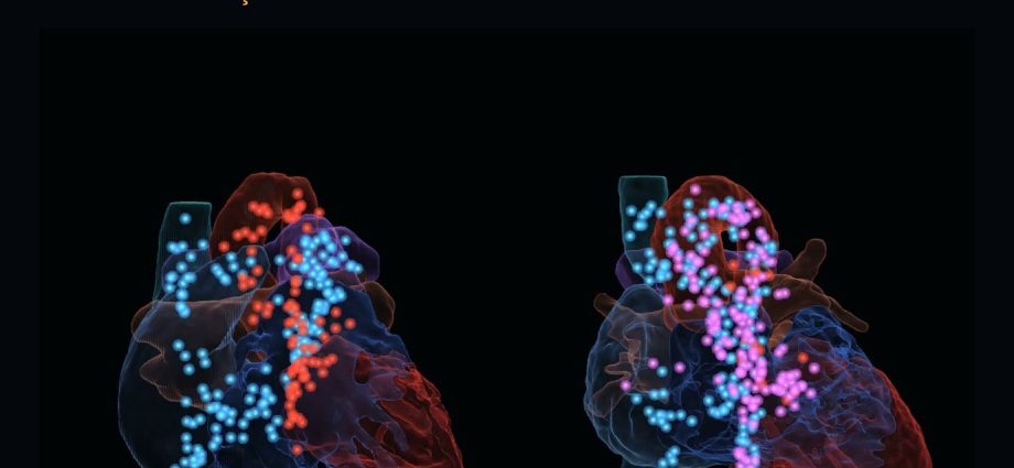
Dois corações reais de crianças, na mesma escala em milímetros, girando lado a lado: um normal e uma transposição. O sangue corre por dentro, pelos caminhos que existem de fato em cada um. Antes de ir ao ar, porém, o vídeo apontava a aorta para o ventrículo errado, e ninguém que não conhecesse a anatomia teria notado. Conto aqui como o filme foi feito e o que a revisão encontrou.
📅 Publicado em 30 de outubro de 2026
Navegue pelo Artigo
Do plantão aos dados, e dos dados ao erro que quase foi ao ar.
- 1. Por que isso importa para quem trabalha na ponta
- 2. Assista: dois corações reais em 73 segundos
- 3. De onde vem o material
- 4. Como o vídeo foi construído
- 5. Os números, sem filtro
- 6. A armadilha: o 3D bonito que ensina errado
- 7. O benefício real para ensinar a transposição
- 8. Cinematic Education: a série começa
- 9. O que fazer na segunda-feira
- 10. Considerações Finais
- 11. Referências
1. Por que isso importa para quem trabalha na ponta
Duas da manhã, UTI cardiológica. Um recém-nascido cianótico chega com o diagnóstico de transposição das grandes artérias. O residente olha o ecocardiograma, ouve “a aorta sai do ventrículo direito” e tenta montar, de cabeça, um objeto tridimensional a partir de cortes planos. A mãe, na porta, ouve a mesma frase e não monta nada.
A transposição é uma cardiopatia espacial. “As artérias estão trocadas” só vira entendimento quando a pessoa enxerga que, no coração normal, a aorta e a pulmonar se cruzam em espiral, enquanto na transposição correm paralelas. No quadro, isso sai torto. Num corte de eco, quase não aparece.
Aqui está o primeiro filme da série Cinematic Education feito com corações reais, junto com os dados de onde ele saiu e os erros que encontrei antes de publicá-lo (foram três, e um deles eu só vi revisando quadro a quadro).
2. Assista: dois corações reais em 73 segundos
Ligue o som. Duas vozes conduzem o filme e a imagem acompanha cada frase.
Coração normal (4 anos) × transposição das grandes artérias com CIV, após cirurgia de Glenn (2 anos). Dados: HVSMR-2.0 (Pace et al., Sci Data 2024) · CC BY 4.0 · modificado. Trilha: “Aphelion” — Scott Buckley · CC BY 4.0.
O que você vai ver
Identificação. As oito estruturas de cada coração — ventrículos, átrios, aorta, pulmonar e as duas veias cavas — rotuladas sobre a parte visível de cada uma.
As grandes artérias. Todo o resto se apaga. De frente, de lado e de cima: no normal, as artérias se enrolam; na transposição, correm paralelas, com a aorta à frente.
O fluxo. Os corações viram vidro e o sangue corre por dentro. Azul é o sangue que volta do corpo; vermelho, o que volta do pulmão. No normal, os circuitos estão em série. Na transposição, o ventrículo direito ejeta na aorta e o sangue azul volta para o corpo.
3. De onde vem o material
Os dois corações vêm do HVSMR-2.0, um conjunto público de 60 ressonâncias cardíacas de pacientes com cardiopatia congênita, publicado em 2024 na Scientific Data por pesquisadores do MIT, do Massachusetts General Hospital e do Boston Children’s Hospital [1]. Cada exame vem com as quatro câmaras e os quatro grandes vasos segmentados manualmente por especialistas, além dos diagnósticos de cada caso.
Os dados estão sob licença Creative Commons Atribuição 4.0 (CC BY 4.0), conferida no próprio registro do repositório: permite uso educacional e comercial, inclusive com modificações, desde que a fonte seja creditada e as modificações indicadas. O crédito aparece no vídeo do início ao fim.
Escolhi dois casos pediátricos: uma criança de 4 anos com coração normal e uma de 2 anos com transposição das grandes artérias (D-TGA), comunicação interventricular e cirurgia de Glenn prévia. Os exames são desidentificados; nenhuma informação que permita reconhecer os pacientes aparece no vídeo ou neste texto.
💡 Tradutor de siglas: o que é uma “segmentação”
A ressonância entrega uma pilha de cortes. Segmentar é marcar, em cada corte, quais pontos pertencem a cada estrutura — este é o ventrículo esquerdo, este é a aorta. Cada ponto do volume é um voxel (o pixel em 3D). No HVSMR-2.0, o que foi marcado é o lúmen: o sangue dentro das cavidades, não o músculo. Por isso o vídeo mostra o “molde” das câmaras e dos vasos, sem parede ventricular e sem septo, e o próprio filme avisa isso na tela.
4. Como o vídeo foi construído
Cada imagem foi calculada a partir dos dados, num motor 3D próprio escrito em Python para a placa de vídeo (ModernGL), com roteiro e decisões guiados pela Metodologia AIMED. Nenhum quadro saiu de gerador de vídeo.
Das máscaras às superfícies
As máscaras viram superfícies 3D pelo algoritmo marching cubes [3], estrutura por estrutura. Os dois corações são reorientados pelo sistema de coordenadas do próprio exame (direita, anterior, superior) e postos na mesma escala em milímetros. A conferência mais boba de todas pegou o primeiro erro: a veia cava superior tem de ficar acima da inferior, e na primeira versão o coração estava de cabeça para baixo.
Os rótulos
Para cada quadro, o motor desenha uma segunda imagem invisível em que cada estrutura tem uma cor-código. Assim ele sabe, pixel a pixel, o que está realmente visível naquele ângulo, e cada rótulo aponta para o ponto mais central da maior região visível da sua estrutura.
O fluxo de sangue
Ninguém desenhou o caminho do sangue. O programa mediu, em cada coração, quais estruturas se tocam de verdade nos dados e, no caso da transposição, encontrou sozinho a aorta saindo do ventrículo direito, a veia cava superior ligada direto à pulmonar (o Glenn) e a comunicação entre os ventrículos, a CIV. Depois traçou cada trajeto pela linha central do lúmen, longe das paredes.
A narração
O diálogo foi escrito a partir das medidas conferidas e gravado com duas vozes sintéticas em português. A fala foi alinhada palavra por palavra ao áudio, e o vídeo foi re-cronometrado pela voz: cada transformação acontece junto com a frase que a explica.
Um exemplo concreto: “o Glenn que o programa achou sozinho”
Eu não disse ao programa que aquele paciente tinha Glenn. Ele mediu: a veia cava superior toca a artéria pulmonar em 191 voxels e o átrio direito em apenas 2. A cava superior drena, portanto, para a pulmonar e não para o átrio, que é a anatomia de uma cirurgia de Glenn (o diagnóstico do conjunto confirma). O fluxo azul do vídeo passa por ali porque o caminho está nos dados.
5. Os números, sem filtro
| Medida (calculada nos dados) | Normal (4 anos) | D-TGA (2 anos) | Fonte | Leitura |
|---|---|---|---|---|
| Raiz da aorta em relação à pulmonar | 13,5 mm atrás, 3,6 mm à direita | 17,6 mm à frente, 11,6 mm à direita | HVSMR-2.0 [1] | ✓ padrão clássico |
| Ventrículo que dá a aorta | VE (244 voxels de contato) | VD (815 voxels de contato) | HVSMR-2.0 [1] | ✓ discordância |
| Veia cava superior drena para | átrio direito (404 voxels) | pulmonar (191) · átrio direito (2) | HVSMR-2.0 [1] | → Glenn |
| Saída do VE para a pulmonar | — | estreita (67 voxels) | HVSMR-2.0 [1] | → dito na tela |
| Linha central do fluxo dentro do lúmen | 99,2% dos pontos | 100% dos pontos | cálculo próprio | ✓ conferido |
💡 Traduzindo para o chão da UTI
A primeira linha é a que o cardiologista pediátrico confere de cabeça: no coração normal, a pulmonar sai à frente e cruza a aorta; na D-TGA, a aorta vem para a frente e para a direita da pulmonar [2]. Os 17,6 mm são a distância medida entre as duas raízes neste coração; é o mesmo número do cartão que aparece no vídeo.
6. A armadilha: o 3D bonito que ensina errado
Um vídeo 3D convence muito mais do que um esquema no quadro. Esse é o benefício, e também o risco: com o rótulo errado, o aluno aprende o errado com mais certeza do que aprenderia com um desenho torto.
A primeira versão deste vídeo tinha três erros, e nenhum deles era visível para quem não conhece a anatomia:
⚠ Os três erros que a revisão encontrou
1. A aorta apontava para o ventrículo direito. No coração normal, o rótulo “Ao” escolhia o ponto da aorta mais próximo da câmera — mas a raiz da aorta fica escondida atrás da via de saída do VD. O ponto caía em cima do ventrículo errado. Percebi revisando o quadro, antes de publicar. A correção trocou a regra: hoje cada rótulo aponta para a parte visível da sua estrutura, verificada pixel a pixel.
2. O sangue era maior que o coração. Os caminhos do fluxo foram calculados numa escala e o coração foi desenhado em outra, cerca de 20% menor. As partículas saíam do lúmen. Hoje os caminhos são guardados em milímetros e a mesma escala é aplicada aos dois.
3. O texto simplificava demais. “Na TGA, os circuitos correm em paralelo” é verdade para a transposição não operada. Este paciente já tinha Glenn. A frase passou a dizer “sem cirurgia, os circuitos ficariam em paralelo”, e a tela agora avisa que a saída do VE para a pulmonar é estreita.
⚠ O que este vídeo não é
As superfícies são o lúmen, não o miocárdio. O fluxo é esquemático: segue as conexões anatômicas reais, mas não é fluxo medido (não há 4D-flow nem simulação de dinâmica de fluidos). São dois casos, longe de cobrir o espectro da transposição. Serve para ensinar; diagnóstico e planejamento cirúrgico ficam fora do alcance dele.
7. O benefício real para ensinar a transposição
O que se ganha é mostrar numa cena só aquilo que o aluno costuma montar aos pedaços, ao longo de semanas de plantão:
A relação espacial. Ver as artérias se enrolando no normal e correndo paralelas na transposição, girando juntas, na mesma escala, responde em segundos a pergunta que o eco 2D deixa no ar.
A conexão. Quem sai de qual ventrículo deixa de ser frase decorada e vira o fluxo azul entrando no VD e saindo pela aorta.
A consequência. Quando o sangue azul volta para o corpo sem passar pelo pulmão, o aluno entende por que o recém-nascido com transposição depende da mistura entre os circuitos , e por que a conduta é urgente.
Para quem isso serve
Residentes de pediatria, cardiologia e terapia intensiva, antes do primeiro plantão de cardiopatia. Enfermagem de UTI cardiológica, para entender por que aquela saturação não sobe com oxigênio. Famílias, numa conversa guiada pela equipe, quando a frase “as artérias estão trocadas” precisa virar algo que se possa ver.
8. Cinematic Education: a série começa
Este é o começo de uma série. Cinematic Education — by Dr. Mbula publica filmes curtos de ensino em 3D, com duas vozes, no mínimo um episódio por semana, sempre com assunto novo. O primeiro episódio usou uma sombra para explicar por que a radiografia de frente engana na ingestão de bateria de botão. Este é o primeiro com corações reais. Antes da série, mostrei aqui um coração 3D didático que pode ser aberto na tela; aquele era geometria simplificada, este é anatomia segmentada de exames reais.
Valem para todos os episódios as mesmas regras: o visual é calculado a partir da física ou dos dados, nunca ilustrado; a tela traz pelo menos um número verificável, saído da mesma conta que move a animação; e o filme declara o que é dado, o que é esquema e de onde veio.
A linha do tempo da série
Da sombra ao coração real, no mínimo um episódio por semana.
9. O que fazer na segunda-feira
Antes de usar qualquer 3D para ensinar
Use IA com método — e com revisão clínica
A Metodologia AIMED ensina médicos a usar inteligência artificial para produzir material de ensino e de trabalho com rigor: fonte primária, conferência e responsabilidade de quem assina.
Considerações Finais
A IA participou do código, das vozes e da animação. Também a pus para revisar o próprio trabalho, e foi assim que apareceram a escala errada do fluxo e a frase simplificada demais. A escolha dos casos, das perguntas, das medidas que valia a pena mostrar e a leitura clínica que pegou a aorta no lugar errado foram trabalho médico. Os dados vieram de especialistas que segmentaram cada exame à mão e os liberaram para que outros pudessem construir em cima.
💡 Connecting the Dots: corações 3D bonitos vão aparecer em toda parte, porque gerador de imagem já faz isso em segundos. Quase ninguém vai mostrar o erro da própria primeira versão, e o meu foi a aorta apontando para o ventrículo errado num vídeo que parecia perfeito. Num filme de ensino, o que vale é a cadeia de conferência entre o dado segmentado por especialistas, o cálculo que respeita a anatomia real e o médico que sabe onde a aorta tem de estar. Sem esse último elo, sobra a aparência do conhecimento. O próximo episódio sai na semana que vem, com outro tema e a mesma regra: número conferido na tela e fonte declarada.
Referências
- Pace DF, Contreras HTM, Romanowicz J, et al. HVSMR-2.0: A 3D cardiovascular MR dataset for whole-heart segmentation in congenital heart disease. Sci Data. 2024;11(1):721. Disponível em: https://doi.org/10.1038/s41597-024-03469-9 · Dados (CC BY 4.0): https://doi.org/10.6084/m9.figshare.c.7074755.v2
- Warnes CA. Transposition of the great arteries. Circulation. 2006;114(24):2699-709. Disponível em: https://doi.org/10.1161/CIRCULATIONAHA.105.592352
- Lorensen WE, Cline HE. Marching cubes: a high resolution 3D surface construction algorithm. ACM SIGGRAPH Computer Graphics. 1987;21(4):163-9. Disponível em: https://doi.org/10.1145/37402.37422
- Buckley S. Aphelion (trilha). Licença CC BY 4.0. Disponível em: https://www.scottbuckley.com.au/library/aphelion/
Transposition of the great arteries in 3D, with real hearts (and the mistake I almost published)
Two real children’s hearts, at the same scale in millimeters, rotating side by side: one normal, one transposition. Blood runs inside them along the paths that actually exist in each. Before release, though, the video pointed the aorta at the wrong ventricle, and nobody who didn’t know the anatomy would have noticed. Here is how the film was made and what the review found.
📅 Published October 30, 2026
Table of Contents
From the bedside to the data, and from the data to the error that almost went out.
- 1. Why this matters at the bedside
- 2. Watch: two real hearts in 73 seconds
- 3. Where the material comes from
- 4. How the video was built
- 5. The numbers, unfiltered
- 6. The trap: beautiful 3D that teaches wrong
- 7. The real benefit for teaching transposition
- 8. Cinematic Education: the series begins
- 9. What to do on Monday
- 10. Final Considerations
- 11. References
1. Why this matters at the bedside
Two in the morning, cardiac ICU. A cyanotic newborn arrives with a diagnosis of transposition of the great arteries. The resident looks at the echocardiogram, hears “the aorta comes off the right ventricle” and tries to build, in their head, a three-dimensional object from flat slices. The mother, at the door, hears the same sentence and builds nothing.
Transposition is a spatial heart defect. “The arteries are switched” only becomes understanding when people see that in the normal heart the aorta and pulmonary artery cross in a spiral, while in transposition they run parallel. On a whiteboard it comes out crooked. In an echo slice it barely shows.
Here is the first Cinematic Education film made with real hearts, along with the data it came from and the errors I found before publishing it (there were three, and I only saw one of them reviewing frame by frame).
2. Watch: two real hearts in 73 seconds
Turn the sound on. Two voices lead the film (narrated in Portuguese, with on-screen captions) and the image follows each sentence.
Normal heart (age 4) × transposition of the great arteries with VSD, after Glenn surgery (age 2). Data: HVSMR-2.0 (Pace et al., Sci Data 2024) · CC BY 4.0 · modified. Music: “Aphelion” — Scott Buckley · CC BY 4.0.
What you will see
Identification. The eight structures of each heart — ventricles, atria, aorta, pulmonary artery and both venae cavae — labeled on the visible part of each one.
The great arteries. Everything else fades out. Front, side and top views: in the normal heart the arteries wrap around each other; in transposition they run parallel, with the aorta in front.
The flow. The hearts turn to glass and blood runs inside. Blue is blood returning from the body; red, blood returning from the lungs. In the normal heart the circuits are in series. In transposition, the right ventricle ejects into the aorta and blue blood goes back to the body.
3. Where the material comes from
Both hearts come from HVSMR-2.0, a public set of 60 cardiac MR scans of patients with congenital heart disease, published in 2024 in Scientific Data by researchers from MIT, Massachusetts General Hospital and Boston Children’s Hospital [1]. Each scan comes with the four chambers and four great vessels manually segmented by experts, plus each case’s diagnoses.
The data are licensed under Creative Commons Attribution 4.0 (CC BY 4.0), checked on the repository record itself: educational and commercial use is allowed, including modifications, as long as the source is credited and changes are indicated. The credit is on screen from start to finish.
I chose two pediatric cases: a 4-year-old with a normal heart and a 2-year-old with transposition of the great arteries (D-TGA), ventricular septal defect and prior Glenn surgery. The scans are de-identified; no information that could identify the patients appears in the video or in this text.
💡 Jargon translator: what is a “segmentation”
MRI delivers a stack of slices. Segmenting means marking, on each slice, which points belong to each structure — this is the left ventricle, this is the aorta. Each point of the volume is a voxel (a 3D pixel). In HVSMR-2.0, what was marked is the lumen: the blood inside the chambers, not the muscle. That is why the video shows a “cast” of the chambers and vessels, with no ventricular wall and no septum, and the film itself warns about that on screen.
4. How the video was built
Every frame was computed from the data, in a custom 3D engine written in Python for the graphics card (ModernGL), with script and decisions guided by the AIMED Methodology. No frame came out of a video generator.
From masks to surfaces
The masks become 3D surfaces through the marching cubes algorithm [3], structure by structure. Both hearts are reoriented by the scan’s own coordinate system (right, anterior, superior) and placed at the same scale in millimeters. The silliest check of all caught the first error: the superior vena cava must sit above the inferior one, and in the first version the heart was upside down.
The labels
For each frame, the engine draws a second, invisible image in which each structure has a color code. That way it knows, pixel by pixel, what is actually visible at that angle, and each label points to the most central point of the largest visible region of its structure.
The blood flow
Nobody drew the blood path. The program measured, in each heart, which structures actually touch in the data and, in the transposition case, found on its own the aorta arising from the right ventricle, the superior vena cava connected directly to the pulmonary artery (the Glenn) and the communication between the ventricles, the VSD. It then traced each path along the centerline of the lumen, away from the walls.
The narration
The dialogue was written from the verified measurements and recorded with two synthetic voices in Portuguese. Speech was aligned word by word to the audio, and the video was retimed by the voice: each transformation happens together with the sentence that explains it.
A concrete example: “the Glenn the program found on its own”
I did not tell the program that this patient had a Glenn. It measured: the superior vena cava touches the pulmonary artery over 191 voxels and the right atrium over only 2. So the superior cava drains into the pulmonary artery and not into the atrium, which is the anatomy of a Glenn operation (the dataset’s diagnosis confirms it). The blue flow in the video goes that way because the path is in the data.
5. The numbers, unfiltered
| Measurement (computed from the data) | Normal (age 4) | D-TGA (age 2) | Source | Reading |
|---|---|---|---|---|
| Aortic root relative to pulmonary root | 13.5 mm behind, 3.6 mm to the right | 17.6 mm in front, 11.6 mm to the right | HVSMR-2.0 [1] | ✓ classic pattern |
| Ventricle giving rise to the aorta | LV (244 contact voxels) | RV (815 contact voxels) | HVSMR-2.0 [1] | ✓ discordance |
| Superior vena cava drains into | right atrium (404 voxels) | pulmonary artery (191) · right atrium (2) | HVSMR-2.0 [1] | → Glenn |
| LV outflow to the pulmonary artery | — | narrow (67 voxels) | HVSMR-2.0 [1] | → stated on screen |
| Flow centerline inside the lumen | 99.2% of points | 100% of points | own calculation | ✓ checked |
💡 Translating to the ICU floor
The first row is the one a pediatric cardiologist checks in their head: in the normal heart, the pulmonary artery arises in front and crosses the aorta; in D-TGA, the aorta comes forward and to the right of the pulmonary artery [2]. The 17.6 mm is the measured distance between the two roots in this heart; it is the same number shown on the video’s card.
6. The trap: beautiful 3D that teaches wrong
A 3D video is far more convincing than a sketch on the board. That is the benefit, and also the risk: with a wrong label, the student learns the wrong thing with more certainty than from a crooked drawing.
The first version of this video had three errors, none of them visible to someone who doesn’t know the anatomy:
⚠ The three errors the review found
1. The aorta pointed at the right ventricle. In the normal heart, the “Ao” label picked the point of the aorta closest to the camera — but the aortic root is hidden behind the RV outflow tract. The point landed on the wrong ventricle. I caught it while reviewing the frame, before publishing. The fix changed the rule: each label now points to the visible part of its structure, checked pixel by pixel.
2. The blood was bigger than the heart. The flow paths were computed at one scale and the heart was drawn at another, about 20% smaller. Particles left the lumen. Paths are now stored in millimeters and the same scale is applied to both.
3. The text oversimplified. “In TGA, the circuits run in parallel” is true for unoperated transposition. This patient already had a Glenn. The sentence now says “without surgery, the circuits would run in parallel”, and the screen now warns that the LV-to-pulmonary outflow is narrow.
⚠ What this video is not
The surfaces are the lumen, not the myocardium. The flow is schematic: it follows the real anatomical connections, but it is not measured flow (no 4D-flow, no computational fluid dynamics). These are two cases, far from covering the spectrum of transposition. It is for teaching; diagnosis and surgical planning are beyond its reach.
7. The real benefit for teaching transposition
What you gain is showing in a single scene what learners usually assemble piece by piece over weeks of shifts:
The spatial relationship. Seeing the arteries wrap around each other in the normal heart and run parallel in transposition, rotating together at the same scale, answers in seconds the question a 2D echo leaves open.
The connection. Which artery comes off which ventricle stops being a memorized sentence and becomes blue flow entering the RV and leaving through the aorta.
The consequence. When blue blood goes back to the body without passing through the lungs, the learner understands why a newborn with transposition depends on mixing between the circuits , and why management is urgent.
Who this is for
Residents in pediatrics, cardiology and critical care, before their first congenital-heart shift. Nurses in the cardiac ICU, to understand why that saturation does not rise with oxygen. Families, in a conversation guided by the team, when “the arteries are switched” needs to become something they can see.
8. Cinematic Education: the series begins
This is the start of a series. Cinematic Education — by Dr. Mbula publishes short 3D teaching films with two voices, at least one episode a week, always on a new topic. The first episode used a shadow to explain why the frontal X-ray misleads in button battery ingestion. This one is the first with real hearts. Before the series, I showed here a teaching 3D heart you can open on screen; that one was simplified geometry, this one is anatomy segmented from real scans.
The same rules apply to every episode: the visual is computed from physics or data, never illustrated; the screen carries at least one verifiable number, taken from the same calculation that drives the animation; and the film declares what is data, what is schematic and where it came from.
The series timeline
From a shadow to a real heart, at least one episode a week.
9. What to do on Monday
Before using any 3D model to teach
Use AI with method — and with clinical review
The AIMED Methodology teaches physicians to use artificial intelligence to produce teaching and working material with rigor: primary sources, verification and accountability of whoever signs.
Final Considerations
AI took part in the code, the voices and the animation. I also had it review its own work, and that is how the wrong flow scale and the oversimplified sentence came up. Choosing the cases, the questions, the measurements worth showing, and the clinical reading that caught the aorta in the wrong place were medical work. The data came from experts who segmented each scan by hand and released them so others could build on top.
💡 Connecting the Dots: beautiful 3D hearts will show up everywhere, because image generators already make them in seconds. Almost nobody will show the error in their own first version, and mine was the aorta pointing at the wrong ventricle in a video that looked perfect. In a teaching film, what counts is the chain of verification between data segmented by experts, a computation that respects real anatomy and a physician who knows where the aorta has to be. Without that last link, all that’s left is the appearance of knowledge. The next episode comes out next week, with a new topic and the same rule: a checked number on screen and a declared source.
References
- Pace DF, Contreras HTM, Romanowicz J, et al. HVSMR-2.0: A 3D cardiovascular MR dataset for whole-heart segmentation in congenital heart disease. Sci Data. 2024;11(1):721. Available at: https://doi.org/10.1038/s41597-024-03469-9 · Data (CC BY 4.0): https://doi.org/10.6084/m9.figshare.c.7074755.v2
- Warnes CA. Transposition of the great arteries. Circulation. 2006;114(24):2699-709. Available at: https://doi.org/10.1161/CIRCULATIONAHA.105.592352
- Lorensen WE, Cline HE. Marching cubes: a high resolution 3D surface construction algorithm. ACM SIGGRAPH Computer Graphics. 1987;21(4):163-9. Available at: https://doi.org/10.1145/37402.37422
- Buckley S. Aphelion (music). CC BY 4.0 license. Available at: https://www.scottbuckley.com.au/library/aphelion/
◆ Novidades
- O coração que eu posso abrir na tela
- Quem Escolhe a Resposta Que a IA Dá ao Seu Residente às Três da Manhã
- A Regra de IA do CFM Também Entra no Consultório — o Artigo 14 Que Ninguém Leu Até o Fim