
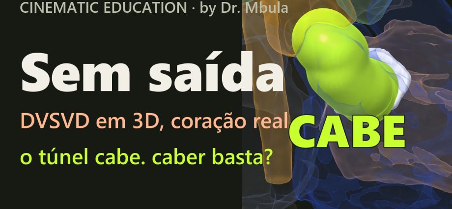
Dupla via de saída do ventrículo direito em 3D: o túnel cabe, mas caber não basta
Na dupla via de saída do ventrículo direito, a aorta e a pulmonar saem do VD, e o sangue do VE só tem uma porta: a CIV. Num coração real de ressonância, um tubo de 13 mm cabe da CIV até a aorta, com 30 mm e nenhum voxel dentro da pulmonar. A CIV, porém, está a 6,3 mm da pulmonar e a 11,9 mm da aorta. A imagem responde à pergunta “dois ventrículos ou um?”. O caminho fica para o time.
📅 Novembro de 2026
Navegue pelo Artigo
Uma pergunta de reunião clínica, um coração real e o que a imagem não consegue decidir.
- 1. A pergunta da reunião
- 2. Assista: Sem Saída
- 3. Passeio 3D em nove passos
- 4. O que é a dupla via de saída do ventrículo direito
- 5. O coração real, em números
- 6. Por que esta CIV não é “remota”
- 7. O teste do tubo de 13 mm
- 8. Duas rotas para dois ventrículos
- 9. O que a imagem não mostra
- 10. Na UTI, depois de cada rota
- 11. Como foi feito e o que deu errado
- 12. O método em 59 corações
- 13. O que é real e o que é calculado
- 14. Perguntas frequentes
- 15. Considerações Finais
- 16. Referências
1. A pergunta da reunião
A cena é ilustrativa, a anatomia é real. Um lactente com dupla via de saída do ventrículo direito já passou pela bandagem da artéria pulmonar, que segura o fluxo para o pulmão e compra tempo. Na reunião clínica de amanhã, alguém vai fazer a pergunta que decide a vida inteira dele: dois ventrículos ou um?
Dois ventrículos significam fechar a CIV de modo que o VE ejete na aorta. Um ventrículo significa o caminho de Glenn e Fontan, sem bomba para o pulmão. A resposta depende de geometria: onde a CIV abre, a que distância das valvas, e se há espaço dentro do VD para levar o sangue do VE até a saída certa.
Fui medir isso num coração real.
2. Assista: Sem Saída
Cerca de 100 segundos, uma voz só. No meio do filme há uma pausa para você votar nos comentários antes de ver a resposta. Depois a câmera entra no ventrículo direito e um tubo cresce da CIV até a aorta, com o contador da pulmonar parado em zero.
3. Passeio 3D em nove passos
Os dois corações reais lado a lado, na mesma escala: um normal, de 17 anos, e o lactente com dupla via de saída do ventrículo direito. Cada passo acende só o que importa: quem sai de cada ventrículo, a CIV, as artérias lado a lado, o tubo de 13 mm e a outra rota. A bússola mostra sempre o lado do paciente, e o último passo é um autoteste.
Dois ventrículos ou um? Decida em nove passos.
Cada passo acende só o que importa nos dois corações reais, lado a lado e na mesma escala. O tubo verde é um cálculo, não anatomia. Depois gire, amplie e faça o teste.
O modelo só é baixado quando você abre.
4. O que é a dupla via de saída do ventrículo direito
Na dupla via de saída do ventrículo direito (DVSVD, ou DORV em inglês), as duas grandes artérias saem inteira ou predominantemente do VD. A nomenclatura internacional de cirurgia congênita pede que cada caso seja descrito por seis itens: a relação da CIV com as valvas semilunares, a relação das grandes artérias entre si, a anatomia coronária, a obstrução das vias de saída, a distância entre a tricúspide e a pulmonar e as lesões associadas [1]. A descontinuidade mitro-arterial e o cone bilateral ajudam, mas não são pré-requisito do diagnóstico [1].
O ponto central é a CIV. Ela é a única saída do VE. Conforme a posição dela, a DVSVD se comporta como uma CIV grande, como uma tetralogia, como uma transposição ou como algo que não se encaixa em nenhuma dessas formas.
Como ler direita e esquerda neste artigo
VD e VE são nomes pela morfologia da câmara. Quando o texto fala de lado, é sempre o lado do paciente. Nas imagens de frente, a direita do paciente aparece à esquerda da imagem, como numa radiografia.

5. O coração real, em números
O caso é o pat48 da base aberta HVSMR-2.0: 60 ressonâncias de cardiopatias congênitas com as câmaras e os vasos segmentados à mão pelos autores [2]. Entre os 22 casos marcados como DVSVD, escolhi este por ter dois ventrículos de tamanho parecido, nenhum artefato de imagem e a paliação ainda antes da decisão. Os planos valvares são as superfícies em que o sangue de uma câmara encosta no da outra, numa grade de 0,7 mm.
| Medida | Valor | Leitura |
|---|---|---|
| Conexões atrioventriculares | AE→VE e AD→VD; zero contato cruzado | concordantes |
| Aorta e pulmonar junto ao VE | 0 voxel | as duas saem do VD |
| Posição das grandes artérias | aorta 19 mm à direita da pulmonar | lado a lado |
| VE / VD (volume do molde) | 0,92 | ventrículos equilibrados |
| Mitral / tricúspide (área) | 0,91 | valvas de entrada equilibradas |
| Raiz aórtica junto à valva | 13,0 mm | medida da RM; não é o anel do eco |
| Tronco da pulmonar | 12,1 → 5,9 mm | estreitamento focal da bandagem |
| CIV → valva aórtica (borda a borda) | 11,9 mm | 0,9 vez a raiz aórtica |
| CIV → valva pulmonar (borda a borda) | 6,3 mm | a CIV está mais perto da pulmonar |
| Tricúspide → valva pulmonar | 19,9 mm | item exigido pela nomenclatura [1] |
| Mitral → valvas aórtica / pulmonar | 15,0 / 12,0 mm | sem continuidade mitro-arterial |
Os volumes são do molde de sangue e não foram indexados pela superfície corporal, que a base não traz. Por isso o artigo usa razões entre os dois lados, sem Z-score.

6. Por que esta CIV não é “remota”
Comecei procurando uma CIV remota, a forma mais difícil da DVSVD. Lacour-Gayet define a CIV não relacionada por três critérios: ela fica a mais de um diâmetro aórtico das duas valvas arteriais, as duas artérias saem inteiras do VD e há cone duplo [3].
Na primeira triagem medi do centro da CIV ao centro da valva aórtica e obtive 24,6 mm, quase duas vezes a raiz. Pareceu remota. O critério, porém, mede de borda a borda. Refeita assim, a distância caiu para 11,9 mm, e a CIV ficou a 6,3 mm da pulmonar. Pelo critério correto, esta CIV não é remota: está relacionada à pulmonar.
O que muda
As artérias lado a lado, com a aorta à direita, e a CIV perto da pulmonar formam o padrão do espectro tipo transposição. Não chamo o caso de Taussig-Bing, porque esse rótulo exige ver o cone, e o músculo não está segmentado. O nome que dá para defender é este: DVSVD com conexão AV concordante, artérias lado a lado e CIV mais próxima da pulmonar, depois de bandagem.
7. O teste do tubo de 13 mm
Para ter dois ventrículos, o sangue do VE precisa chegar à aorta. Desenhei dentro do VD o caminho mais curto da CIV até a valva aórtica, afastado das paredes, e o transformei num tubo de 13 mm, a largura da raiz aórtica junto à valva.
| Teste | Resultado |
|---|---|
| Comprimento da linha central | 30,3 mm |
| Voxels do tubo dentro da pulmonar | 0 |
| Voxels do tubo fora do sangue | 55 (0,4%) |
| Fração do VD ocupada | 11% |
| Saída VD→pulmonar, maior esfera que passa | 11,5 mm com o tubo, 11,5 mm sem ele |
| Menor distância do trajeto à valva pulmonar / à tricúspide | 12,5 / 10,6 mm |
O tubo cabe. Não atravessa parede, não entra na pulmonar e não estreita a saída do VD. No filme, o contador da pulmonar fica em zero enquanto o tubo cresce, e esse número sai da mesma máscara que gera o tubo na tela.

8. Duas rotas para dois ventrículos
Caber não é o mesmo que ser a melhor cirurgia. Quando a CIV está perto da pulmonar, existe outra rota: fechar a CIV para a pulmonar e trocar as artérias de lugar, com a cirurgia de Jatene. Masuda e colaboradores operaram assim 27 pacientes com DVSVD e CIV subpulmonar quando o túnel intraventricular não era viável. Houve um óbito operatório. Nos pacientes com artérias lado a lado, a coronária esquerda passava atrás da pulmonar em 10 de 12, e 9 dos 27 tinham anomalia do arco [4].
No outro extremo, na CIV não relacionada, Li e colaboradores compararam 40 pacientes com o túnel até a aorta e 35 com o túnel até a pulmonar mais a troca arterial. As obstruções do túnel apareceram só no primeiro grupo: 10 contra 0 [5]. O caso deste artigo não é desse tipo, mas o dado mostra por que um túnel longo até a aorta preocupa o cirurgião.
Na série de Tchervenkov, 24 lactentes com DVSVD foram corrigidos com quatro técnicas diferentes, escolhidas caso a caso [6]. Esse é o padrão da doença: a rota é individual.

9. O que a imagem não mostra
Pelos dados desta ressonância, a resposta é dois ventrículos: câmaras e valvas de entrada equilibradas, uma CIV do tamanho da valva aórtica (razão de área 0,87) e espaço para um tubo sem fechar a saída do VD. A escolha da rota depende do que esta imagem não mostra:
- O cone e o septo infundibular: o músculo da saída decide se o túnel fica livre ou obstruído.
- As cordas da tricúspide: podem atravessar o caminho do túnel.
- As coronárias: decidem o risco da troca arterial.
- O gradiente da bandagem e a resistência vascular pulmonar: fisiologia, não anatomia.
Quem decide é o time: cardiologista, cirurgião e intensivista, com eco, cateterismo quando indicado e a criança na frente.
10. Na UTI, depois de cada rota
Esta seção é a conduta que eu levo para o plantão, não um dado do estudo.
- Depois da bandagem: a saturação e o eco dizem se a faixa está apertada demais (cianose) ou frouxa (hiperfluxo pulmonar). A bandagem compra tempo, mas não corrige.
- Depois do túnel até a aorta: procurar obstrução da via de saída do VE e CIV residual. Um túnel longo tem mais motivo para obstruir [5].
- Depois da troca arterial: isquemia coronária vem primeiro. Olhar o ECG seriado, o lactato e a função ventricular nas primeiras horas, e saber de antemão a anatomia coronária descrita na cirurgia [4].
- Nas duas: arritmias pós-operatórias e bloqueio AV, porque a sutura passa perto do sistema de condução.
11. Como foi feito e o que deu errado
Usei a segmentação feita à mão da base HVSMR-2.0 [2] e escrevi scripts que medem contatos, distâncias e trajetos. O filme sai de um motor 3D próprio, e cada número na tela vem da mesma conta que move a imagem.
- Erro 1, o maior: chamei a CIV de remota medindo de centro a centro (24,6 mm). De borda a borda deu 11,9 mm. O caso continuou bom; o rótulo estava errado. Por isso o vídeo não diz “remota”.
- Erro 2: medi a bandagem em faixas de 3 mm ao longo do tronco e obtive 7,0 mm. Em faixas de 1 mm, que localizam melhor o ponto mais estreito, deu 5,9 mm. Ficou o segundo valor, com o método escrito.
- Erro 3: no inglês, a voz sintética repetiu “or one” fora do roteiro. Cortei a repetição; o resto da fala ficou intacto, em velocidade normal.
- Revisão: fiz uma revisão crítica no papel de um ecocardiografista sênior, simulada com IA. Ela trocou “anel” por “raiz junto à valva”, tirou “CIV não restritiva” e “perimembranosa” e transformou a linha do túnel no tubo de 13 mm. Ainda não é a revisão de um cardiologista de carne e osso.
12. O método em 59 corações
Antes de confiar no método, testei-o na base inteira. Escrevi as regras antes de ver os resultados e não as ajustei depois. Comparei o diagnóstico derivado automaticamente da segmentação com o diagnóstico registrado em cada caso.
| Critério | Sensibilidade | Especificidade |
|---|---|---|
| CIV | 0,97 | 0,77 |
| DVSVD | 0,82 | 0,86 |
| Ventrículo único | 1,00 | 0,96 |
| Bandagem da pulmonar | 0,83 | 1,00 |
| Fontan | 0,89 | 1,00 |
| Veia cava superior bilateral | 1,00 | 1,00 |
| Dextrocardia | 0,90 | 1,00 |
| CIA ou átrio comum | 0,62 | 0,63 |
| Transposição não operada | 0,40 | 1,00 |
| Anastomose aorta-pulmonar | 0,00 | 0,89 |
| Total (59 casos × 12 critérios) | 636 de 708 decisões batem (89,8%); acurácia balanceada média de 82% | |
Os erros têm padrão. O método falha onde a parede é mais fina que o pixel: o septo atrial e os vasos que encostam um no outro viram falsas comunicações. Também falha na atresia pulmonar, em que transposição e DVSVD ficam indistinguíveis sem a conexão da pulmonar. O pat48 está do lado confiável: a CIV tem 269 mm² de contato, acima de qualquer falso positivo comparável, e as duas artérias saem do VD com conexões amplas.

13. O que é real e o que é calculado
Limites
Real: a anatomia do pat48, a segmentação dos autores da base e o corte de ressonância. Calculado: distâncias, volumes, o tubo e a validação, por scripts próprios sobre a segmentação. Esquema: a troca das artérias no filme e as partículas da via de saída. Ilustrativo: a história clínica, a reunião e a idade descrita. O método roda sobre a segmentação feita por especialistas, não sobre a imagem crua, e não substitui o eco nem o especialista. Material educacional, sem uso diagnóstico.
14. Perguntas frequentes
Esta é uma CIV não relacionada?
Não. Medida de borda a borda, ela fica a 11,9 mm da aorta e a 6,3 mm da pulmonar. O critério de não relacionada exige mais de um diâmetro aórtico das duas valvas [3].
Então é um Taussig-Bing?
O padrão aponta para o espectro tipo transposição, mas confirmar o nome exige ver o cone, que não está segmentado. O artigo não usa o rótulo.
Se o tubo cabe, por que não operar assim?
Porque caber é geometria. A escolha depende do músculo da saída, das cordas e das coronárias, que esta imagem não mostra, e da experiência do serviço [4–6].
A IA fez o diagnóstico?
Não. A segmentação é manual, feita pelos autores da base. Os scripts medem o que a segmentação mostra e foram testados nos 59 casos, com os erros expostos.
Quer ensinar anatomia assim?
A Metodologia AIMED organiza o uso de IA em medicina, com revisão clínica em cada etapa.
15. Considerações Finais
A pergunta “dois ventrículos ou um?” costuma ser respondida no eco e na reunião. Aqui, a ressonância pública de um lactente respondeu metade dela: há dois ventrículos equilibrados e espaço para o túnel. A outra metade, que é o caminho, depende do que a imagem não mostra.
💡 Connecting the Dots. O erro de medir do centro, e não da borda, transformou uma CIV comum em “remota”. É o mesmo erro que se comete à beira do leito quando se descreve a CIV pelo que parece no 3D, e não pelo que a definição mede. O método não é perfeito, mas mostra até onde dá para chegar com dados abertos, contas que qualquer colega pode repetir e erros escritos.
Leia também o episódio anterior, Transposição corrigida em 3D, com o mesmo método aplicado a outra cardiopatia.
Este coração só pôde ser estudado porque o exame de alguém está em uma base pública.
Referências
- Walters HL, Mavroudis C, Tchervenkov CI, Jacobs JP, Lacour-Gayet F, Jacobs ML. Congenital Heart Surgery Nomenclature and Database Project: double outlet right ventricle. Ann Thorac Surg. 2000;69(4 Suppl):S249-63. PMID: 10798433. DOI: 10.1016/s0003-4975(99)01247-3
- Pace DF, Contreras HTM, Romanowicz J, et al. HVSMR-2.0: A 3D cardiovascular MR dataset for whole-heart segmentation in congenital heart disease. Sci Data. 2024;11:721. PMID: 38956063. DOI: 10.1038/s41597-024-03469-9
- Lacour-Gayet F. Biventricular repair of double outlet right ventricle with noncommitted ventricular septal defect. Semin Thorac Cardiovasc Surg Pediatr Card Surg Annu. 2002;5:163-72. PMID: 11994877. DOI: 10.1053/pcsu.2002.31491
- Masuda M, Kado H, Shiokawa Y, et al. Clinical results of arterial switch operation for double-outlet right ventricle with subpulmonary VSD. Eur J Cardiothorac Surg. 1999;15(3):283-8. PMID: 10333024. DOI: 10.1016/s1010-7940(99)00002-0
- Li S, Ma K, Hu S, et al. Biventricular repair for double outlet right ventricle with non-committed ventricular septal defect. Eur J Cardiothorac Surg. 2015;48(4):580-7. PMID: 25612745. DOI: 10.1093/ejcts/ezu523
- Tchervenkov CI, Marelli D, Béland MJ, et al. Institutional experience with a protocol of early primary repair of double-outlet right ventricle. Ann Thorac Surg. 1995;60(6 Suppl):S610-3. PMID: 8604947. DOI: 10.1016/0003-4975(95)00855-1
Música do filme: ‘In This Moment’ by Scott Buckley – released under CC-BY 4.0. www.scottbuckley.com.au
Double outlet right ventricle in 3D: the tunnel fits, but fitting is not enough
In double outlet right ventricle, both the aorta and the pulmonary artery leave the RV, and the LV has a single door: the VSD. In a real MRI heart, a 13 mm tube fits from the VSD to the aorta: 30 mm long, with no voxel inside the pulmonary artery. But the VSD lies 6.3 mm from the pulmonary valve and 11.9 mm from the aortic one. The image answers “two ventricles or one?”. The route is the team’s call.
📅 November 2026
Navigate the Article
A case-conference question, a real heart, and what the image cannot decide.
- 1. The conference question
- 2. Watch: No Way Out
- 3. Nine-step 3D tour
- 4. What double outlet right ventricle is
- 5. The real heart, in numbers
- 6. Why this VSD is not “remote”
- 7. The 13 mm tube test
- 8. Two routes to two ventricles
- 9. What the image does not show
- 10. In the ICU, after each route
- 11. How it was made and what went wrong
- 12. The method on 59 hearts
- 13. What is real and what is computed
- 14. Frequently asked questions
- 15. Final Thoughts
- 16. References
1. The conference question
The scene is illustrative; the anatomy is real. An infant with double outlet right ventricle has already had a pulmonary artery band, which limits lung blood flow and buys time. At tomorrow’s case conference, someone will ask the question that shapes the rest of the child’s life: two ventricles or one?
Two ventricles means closing the VSD so that the LV ejects into the aorta. One ventricle means the Glenn and Fontan path, with no pump to the lungs. The answer is geometry: where the VSD opens, how far it is from each valve, and whether the RV has room to carry LV blood to the right exit.
I went to measure that in a real heart.
2. Watch: No Way Out
About 90 seconds, one voice. Halfway through, there is a pause for you to vote in the comments before the answer. Then the camera goes into the right ventricle and a tube grows from the VSD to the aorta while the pulmonary counter stays at zero.
3. Nine-step 3D tour
The two real hearts side by side, at the same scale: a normal one, age 17, and the infant with double outlet right ventricle. Each step lights only what matters: what leaves each ventricle, the VSD, the side-by-side arteries, the 13 mm tube and the other route. The compass always shows the patient’s side, and the last step is a self-test.
Two ventricles or one? Decide in nine steps.
Each step lights only what matters in the two real hearts, side by side at the same scale. The green tube is a calculation, not anatomy. Then rotate, zoom and take the test.
The model downloads only when you open it.
4. What double outlet right ventricle is
In double outlet right ventricle (DORV), both great arteries arise entirely or predominantly from the RV. The international congenital heart surgery nomenclature asks for six items in every case: the VSD’s relation to the semilunar valves, the relation of the great arteries to each other, the coronary anatomy, outflow tract obstruction, the tricuspid-to-pulmonary distance and associated lesions [1]. Mitral-arterial discontinuity and a bilateral conus help, but neither is required for the diagnosis [1].
The VSD is the key. It is the LV’s only exit. Depending on where it lies, DORV behaves like a large VSD, like tetralogy, like transposition, or like none of them.
How to read right and left in this article
RV and LV are named by chamber morphology. Whenever the text mentions a side, it is the patient’s side. In front views, the patient’s right appears on the left of the image, as in a chest X-ray.

5. The real heart, in numbers
The case is pat48 from the open HVSMR-2.0 dataset: 60 MRI scans of congenital heart disease, with chambers and vessels segmented by hand by the authors [2]. Of the 22 cases labelled DORV, I picked this one because it has two similar-sized ventricles, no image artefact, and palliation still short of the final decision. Valve planes are the surfaces where blood of one chamber touches blood of the next, on a 0.7 mm grid.
| Measure | Value | Reading |
|---|---|---|
| Atrioventricular connections | LA→LV and RA→RV; zero cross-contact | concordant |
| Aorta and pulmonary touching the LV | 0 voxels | both arise from the RV |
| Great artery position | aorta 19 mm to the right of the pulmonary | side by side |
| LV / RV (cast volume) | 0.92 | balanced ventricles |
| Mitral / tricuspid (area) | 0.91 | balanced inflow valves |
| Aortic root at the valve | 13.0 mm | MRI measure; not the echo annulus |
| Pulmonary trunk | 12.1 → 5.9 mm | focal narrowing from the band |
| VSD → aortic valve (edge to edge) | 11.9 mm | 0.9 times the aortic root |
| VSD → pulmonary valve (edge to edge) | 6.3 mm | the VSD is closer to the pulmonary |
| Tricuspid → pulmonary valve | 19.9 mm | item required by the nomenclature [1] |
| Mitral → aortic / pulmonary valves | 15.0 / 12.0 mm | no mitral-arterial continuity |
Volumes are blood-cast volumes and are not indexed to body surface area, which the dataset does not provide. The article therefore uses side-to-side ratios, without Z-scores.

6. Why this VSD is not “remote”
I started out looking for a remote VSD, the hardest form of DORV. Lacour-Gayet defines a noncommitted VSD by three criteria: it lies more than one aortic diameter from both arterial valves, both arteries arise entirely from the RV, and there is a double conus [3].
In the first screen I measured from the centre of the VSD to the centre of the aortic valve and got 24.6 mm, almost twice the root. It looked remote. The criterion, however, measures edge to edge. Measured that way, the distance dropped to 11.9 mm, and the VSD sat 6.3 mm from the pulmonary valve. By the correct criterion, this VSD is not remote: it is related to the pulmonary valve.
What changes
Side-by-side arteries with the aorta on the right and a VSD near the pulmonary valve make up the transposition-type pattern. I do not call it Taussig-Bing, because that label requires seeing the conus, and muscle is not segmented. The name I can defend is: DORV with concordant AV connections, side-by-side arteries and a VSD closer to the pulmonary valve, after banding.
7. The 13 mm tube test
For two ventricles, LV blood must reach the aorta. Inside the RV I traced the shortest path from the VSD to the aortic valve, kept away from the walls, and turned it into a 13 mm tube, the width of the aortic root at the valve.
| Test | Result |
|---|---|
| Centreline length | 30.3 mm |
| Tube voxels inside the pulmonary artery | 0 |
| Tube voxels outside the blood | 55 (0.4%) |
| Share of the RV occupied | 11% |
| RV→pulmonary outflow, largest sphere that passes | 11.5 mm with the tube, 11.5 mm without it |
| Closest approach to the pulmonary valve / tricuspid | 12.5 / 10.6 mm |
The tube fits. It crosses no wall, does not enter the pulmonary artery and does not narrow the RV outflow. In the film, the pulmonary counter stays at zero while the tube grows, and that number comes from the same mask that draws the tube on screen.

8. Two routes to two ventricles
Fitting is not the same as being the best operation. When the VSD sits near the pulmonary valve, there is another route: close the VSD to the pulmonary valve and swap the arteries with an arterial switch. Masuda and colleagues operated on 27 patients with DORV and subpulmonary VSD this way when an intraventricular tunnel was not feasible. There was one operative death. In side-by-side arteries, the left coronary ran behind the pulmonary artery in 10 of 12, and 9 of the 27 had an arch anomaly [4].
At the other end, in noncommitted VSD, Li and colleagues compared 40 patients with a tunnel to the aorta and 35 with a tunnel to the pulmonary artery plus switch. Tunnel obstruction appeared only in the first group: 10 versus 0 [5]. This article’s case is not of that type, but the data show why a long tunnel to the aorta worries surgeons.
In Tchervenkov’s series, 24 infants with DORV were repaired with four different techniques, chosen case by case [6]. That is the rule in this disease: the route is individual.

9. What the image does not show
From this MRI, the answer is two ventricles: balanced chambers and inflow valves, a VSD the size of the aortic valve (area ratio 0.87), and room for a tube without closing the RV outflow. Choosing the route depends on what this image does not show:
- The conus and infundibular septum: outflow muscle decides whether the tunnel stays open.
- The tricuspid chords: they may cross the tunnel’s path.
- The coronaries: they set the risk of the arterial switch.
- The band gradient and pulmonary vascular resistance: physiology, not anatomy.
The team decides: cardiologist, surgeon and intensivist, with echo, catheterisation when indicated, and the child in front of them.
10. In the ICU, after each route
This is what I take to the bedside, not a study finding.
- After banding: saturation and echo tell you whether the band is too tight (cyanosis) or too loose (pulmonary overflow). The band buys time; it does not repair.
- After a tunnel to the aorta: look for LV outflow obstruction and residual VSD. A long tunnel has more reason to obstruct [5].
- After an arterial switch: coronary ischaemia first. Watch serial ECGs, lactate and ventricular function in the first hours, and know in advance the coronary anatomy described at surgery [4].
- After either: postoperative arrhythmias and AV block, because the suture line runs close to the conduction system.
11. How it was made and what went wrong
I used the hand-made segmentation of the HVSMR-2.0 dataset [2] and wrote scripts that measure contacts, distances and paths. The film comes from my own 3D engine, and every on-screen number comes from the same calculation that moves the image.
- Error 1, the biggest: I called the VSD remote after measuring centre to centre (24.6 mm). Edge to edge it was 11.9 mm. The case stayed good; the label was wrong. That is why the video never says “remote”.
- Error 2: I measured the band in 3 mm slabs along the trunk and got 7.0 mm. In 1 mm slabs, which locate the narrowest point better, it was 5.9 mm. The second value stayed, with the method written down.
- Error 3: in English, the synthetic voice repeated “or one” off script. I cut the repetition; the rest of the speech is intact, at normal speed.
- Review: I ran a critical review in the role of a senior echocardiographer, simulated with AI. It swapped “annulus” for “root at the valve”, removed “non-restrictive VSD” and “perimembranous”, and turned the tunnel line into the 13 mm tube. It is not yet a review by a flesh-and-blood cardiologist.
12. The method on 59 hearts
Before trusting the method, I tested it on the whole dataset. I wrote the rules before seeing the results and did not tune them afterwards. I compared the diagnosis derived automatically from the segmentation with the diagnosis recorded for each case.
| Criterion | Sensitivity | Specificity |
|---|---|---|
| VSD | 0.97 | 0.77 |
| DORV | 0.82 | 0.86 |
| Single ventricle | 1.00 | 0.96 |
| Pulmonary artery band | 0.83 | 1.00 |
| Fontan | 0.89 | 1.00 |
| Bilateral superior vena cava | 1.00 | 1.00 |
| Dextrocardia | 0.90 | 1.00 |
| ASD or common atrium | 0.62 | 0.63 |
| Unoperated transposition | 0.40 | 1.00 |
| Aorto-pulmonary anastomosis | 0.00 | 0.89 |
| Total (59 cases × 12 criteria) | 636 of 708 decisions match (89.8%); mean balanced accuracy 82% | |
The errors follow a pattern. The method fails where the wall is thinner than a pixel: the atrial septum and vessels lying against each other turn into false communications. It also fails in pulmonary atresia, where transposition and DORV cannot be told apart without the pulmonary connection. Pat48 sits on the reliable side: its VSD has 269 mm² of contact, above any comparable false positive, and both arteries arise from the RV with wide connections.

13. What is real and what is computed
Limits
Real: pat48’s anatomy, the dataset authors’ segmentation and the MRI slice. Computed: distances, volumes, the tube and the validation, by my own scripts on top of the segmentation. Schematic: the arterial swap in the film and the outflow particles. Illustrative: the clinical story, the conference and the age described. The method runs on an expert segmentation, not on the raw image, and is no substitute for echo or the specialist. Educational material, not for diagnostic use.
14. Frequently asked questions
Is this a noncommitted VSD?
No. Measured edge to edge, it lies 11.9 mm from the aorta and 6.3 mm from the pulmonary valve. Noncommitted requires more than one aortic diameter from both valves [3].
So is it Taussig-Bing?
The pattern points to the transposition-type spectrum, but confirming the name requires seeing the conus, which is not segmented. The article does not use the label.
If the tube fits, why not operate that way?
Because fitting is geometry. The choice depends on outflow muscle, chords and coronaries, which this image does not show, and on the unit’s experience [4–6].
Did AI make the diagnosis?
No. The segmentation is manual, by the dataset authors. The scripts measure what the segmentation shows and were tested on the 59 cases, with the errors shown.
Want to teach anatomy like this?
The AIMED Methodology organises the use of AI in medicine, with clinical review at every step.
15. Final Thoughts
“Two ventricles or one?” is usually answered by echo and at the conference table. Here, an infant’s public MRI answered half of it: there are two balanced ventricles and room for the tunnel. The other half, the route, depends on what the image cannot show.
💡 Connecting the Dots. Measuring from the centre instead of the edge turned an ordinary VSD into a “remote” one. It is the same mistake made at the bedside when a VSD is described by how it looks in 3D rather than by what the definition measures. The method is not perfect, but it shows how far you can go with open data, calculations any colleague can repeat, and errors written down.
See also the previous episode, Corrected transposition in 3D, with the same method applied to another defect.
This heart could only be studied because someone’s scan is in a public dataset.
References
- Walters HL, Mavroudis C, Tchervenkov CI, Jacobs JP, Lacour-Gayet F, Jacobs ML. Congenital Heart Surgery Nomenclature and Database Project: double outlet right ventricle. Ann Thorac Surg. 2000;69(4 Suppl):S249-63. PMID: 10798433. DOI: 10.1016/s0003-4975(99)01247-3
- Pace DF, Contreras HTM, Romanowicz J, et al. HVSMR-2.0: A 3D cardiovascular MR dataset for whole-heart segmentation in congenital heart disease. Sci Data. 2024;11:721. PMID: 38956063. DOI: 10.1038/s41597-024-03469-9
- Lacour-Gayet F. Biventricular repair of double outlet right ventricle with noncommitted ventricular septal defect. Semin Thorac Cardiovasc Surg Pediatr Card Surg Annu. 2002;5:163-72. PMID: 11994877. DOI: 10.1053/pcsu.2002.31491
- Masuda M, Kado H, Shiokawa Y, et al. Clinical results of arterial switch operation for double-outlet right ventricle with subpulmonary VSD. Eur J Cardiothorac Surg. 1999;15(3):283-8. PMID: 10333024. DOI: 10.1016/s1010-7940(99)00002-0
- Li S, Ma K, Hu S, et al. Biventricular repair for double outlet right ventricle with non-committed ventricular septal defect. Eur J Cardiothorac Surg. 2015;48(4):580-7. PMID: 25612745. DOI: 10.1093/ejcts/ezu523
- Tchervenkov CI, Marelli D, Béland MJ, et al. Institutional experience with a protocol of early primary repair of double-outlet right ventricle. Ann Thorac Surg. 1995;60(6 Suppl):S610-3. PMID: 8604947. DOI: 10.1016/0003-4975(95)00855-1
Film music: ‘In This Moment’ by Scott Buckley – released under CC-BY 4.0. www.scottbuckley.com.au
◆ Novidades
- Transposição corrigida em 3D: duas trocas que se anulam e o ventrículo que paga a contaNOVO
- Anatomia do olho em 3D: faça o teste do olhinho em um olho realNOVO
- Anatomia da cabeça em 3D: rosto, crânio e cérebro de uma só pessoa