
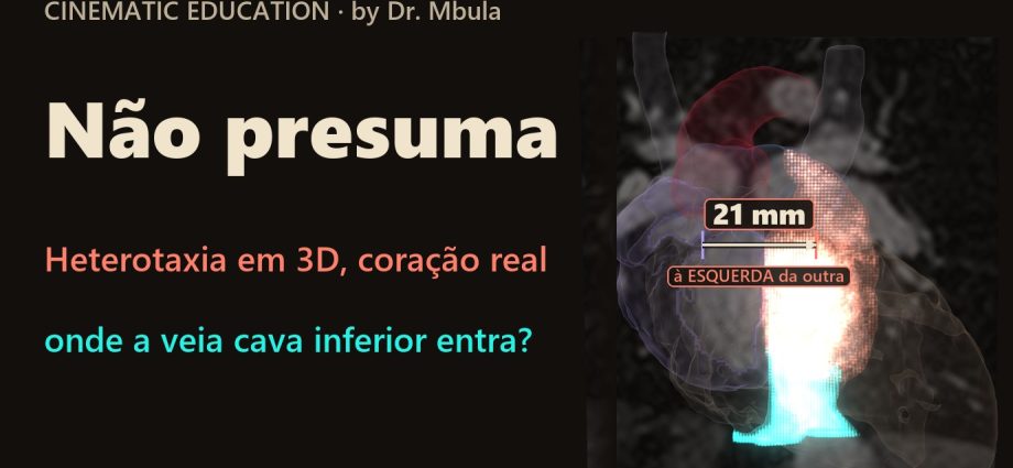
Heterotaxia em 3D: a veia cava inferior entrou do outro lado, e isso não diz qual átrio é qual
Num coração real de ressonância, de um lactente de 1 ano com heterotaxia e sem cirurgia, a veia cava inferior entra na câmara atrial que fica 21 mm à esquerda da outra. No coração de referência, ela entra na câmara da direita, a 19 mm. A tentação é chamar a câmara de “átrio esquerdo”. Na heterotaxia, quem define o átrio é a forma do apêndice, e esta imagem não mostra o apêndice. Cada conexão é procurada, nunca presumida.
📅 Novembro de 2026
Navegue pelo Artigo
Uma pergunta simples de passagem de plantão, um coração real e o que a imagem não consegue nomear.
- 1. A pergunta da passagem de plantão
- 2. Assista: Não Presuma
- 3. Passeio 3D em oito passos
- 4. O que é heterotaxia
- 5. O coração real, em números
- 6. A junção, de perto
- 7. Qual átrio é qual?
- 8. As duas veias cavas superiores
- 9. Rótulo não é anatomia
- 10. O que a imagem não mostra
- 11. Na UTI, antes de presumir
- 12. Como foi feito e o que deu errado
- 13. O que é real e o que é calculado
- 14. Perguntas frequentes
- 15. Considerações Finais
- 16. Referências
1. A pergunta da passagem de plantão
A cena é ilustrativa, a anatomia é real. Um lactente de 1 ano, cianótico. O eco fala em heterotaxia: os órgãos não seguem o lado de sempre. Amanhã a residente apresenta a anatomia para a cirurgia, e eu faço a pergunta mais simples que existe: onde a veia cava inferior entra?
“Na direita. Sempre na direita.” É a resposta certa para quase todos os corações que a gente vê. Na heterotaxia, é exatamente a resposta que não se pode dar sem olhar.
Fui olhar num coração real.
2. Assista: Não Presuma
Cerca de 95 segundos, duas vozes: o ecocardiografista e a residente. O primeiro coração mostra o caminho habitual da veia. No segundo, a câmera acende só o brilho real do sangue na ressonância e sobe com ele, até a veia entrar do outro lado. No fim, os bastidores: onde quase erramos.
3. Passeio 3D em oito passos
Os dois corações reais lado a lado, na mesma escala em milímetros: o de referência, de 4 anos, e o lactente com heterotaxia. Primeiro só a veia cava inferior acende e você tenta adivinhar para onde ela vai. Depois vêm a resposta medida, a junção de perto, as duas cavas superiores e o último passo, um autoteste. A bússola mostra sempre o lado do paciente, e nenhuma imagem está espelhada. Toque em qualquer estrutura para ver o nome.
Onde entra a veia cava inferior? Descubra em oito passos.
Dois corações reais, lado a lado e na mesma escala. Cada passo acende só o que importa; a veia e a câmara que a recebe pulsam de luz. Depois gire, amplie e faça o teste.
O modelo só é baixado quando você abre.
4. O que é heterotaxia
Heterotaxia é o arranjo anormal dos órgãos ao longo do eixo direita-esquerda do corpo: nem o arranjo habitual, nem a imagem em espelho dele. O coração costuma vir junto, com conexões venosas, atriais e ventriculares fora do padrão. A nomenclatura internacional reuniu as definições numa revisão de consenso, que compara a escola europeia de análise segmentar sequencial e a escola de Boston [1].
Para quem cuida dessas crianças, a consequência prática é uma só: as regras de bolso deixam de valer. A veia cava inferior pode entrar na câmara da esquerda, pode ser interrompida, e pode haver duas veias cavas superiores. Nada pode ser deduzido pelo lado. Tudo precisa ser visto.
Como ler direita e esquerda neste artigo
Quando o texto fala de lado, é sempre o lado do paciente. Nas imagens de frente, a direita do paciente aparece à esquerda da imagem, como numa radiografia. As câmaras atriais do coração com heterotaxia não recebem os nomes “direito” e “esquerdo”: são a “câmara que recebe a veia cava inferior” e a “outra câmara atrial”. A seção 7 explica por quê.

5. O coração real, em números
O caso é o pat38 da base aberta HVSMR-2.0: 60 ressonâncias de cardiopatias congênitas com câmaras e vasos segmentados à mão pelos autores [3]. Entre os 13 corações com heterotaxia da base, escolhi um de 1 ano, ainda sem cirurgia, com a junção da veia cava inferior dentro do campo da imagem. O coração de referência é o pat5, de 4 anos. As distâncias são medidas entre os centros das duas câmaras atriais, no eixo direita-esquerda do paciente. O contato é contado em voxels que se tocam face a face.
| Medida | Heterotaxia (1 ano) | Referência (4 anos) |
|---|---|---|
| Câmara que recebe a veia cava inferior | 21,2 mm à esquerda da outra | 18,7 mm à direita da outra |
| Contato da veia cava inferior com a câmara receptora | 312 voxels | 609 voxels |
| Contato da veia cava inferior com a outra câmara | 0 | 0 |
| Veias cavas superiores | duas, uma de cada lado | uma |
| Cava superior esquerda: contato com a câmara receptora / com a outra | 81 / 0 voxels | não se aplica |
| Cava superior direita: contato com a câmara receptora / com a outra | 0 / 57 voxels | não se aplica |
| Veia cava inferior abaixo do fígado | dois ramos sem identidade definida, que se unem dentro do fígado | um tronco |
A base registra para este coração, entre outros achados, heterotaxia, comunicação interatrial e interventricular, dupla via de saída do ventrículo direito e veia cava superior bilateral [3]. O artigo só afirma o que as medidas acima sustentam.

6. A junção, de perto
Um modelo 3D bonito pode enganar. Superfícies suavizadas podem encostar onde a anatomia não encosta. Por isso a junção foi conferida nos cortes originais da ressonância, nos três planos: coronal, sagital e axial. Nos três, a luz da veia cava inferior chega direto à câmara receptora, numa faixa contínua de 312 voxels de contato. Com a outra câmara, o contato é zero.
No filme, a “luz” que sobe pela veia não é desenho. É a intensidade real do sinal da ressonância, em que o sangue brilha, mostrada só dentro da veia e da câmara. O que acende na tela é o que o exame registrou.
O que o contato prova, e o que não prova
Contato face a face entre o sangue da veia e o sangue da câmara mostra a conexão anatômica. Ele não mede fluxo, nem velocidade, nem pressão. Para isso existem o Doppler e o cateterismo.
7. Qual átrio é qual?
A pergunta seguinte é inevitável: se a veia cava inferior entra na câmara da esquerda, então essa câmara é o átrio esquerdo? Não dá para dizer.
Na análise segmentar sequencial, o átrio é identificado pela morfologia do seu apêndice, porque o apêndice é a parte mais constante da câmara. As veias que chegam podem variar, e na heterotaxia variam. Tremblay e colaboradores, ao estudar 188 corações de autópsia com isomerismo, concluíram que o isomerismo é mais bem separado pela morfologia dos apêndices atriais [2]. Na mesma série, a interrupção da veia cava inferior se associou ao isomerismo esquerdo [2].
A segmentação desta base é do sangue e não separa os apêndices. Sem ver o apêndice, chamar a câmara receptora de “átrio esquerdo” ou de “átrio direito” seria um palpite com cara de laudo. Por isso o filme, o passeio 3D e este artigo usam os nomes do que foi medido.

Uma nota para o especialista
O critério “a forma do apêndice define o átrio” é o da análise segmentar sequencial, a escola europeia associada a Anderson. A escola de Boston, de Van Praagh, descreve a heterotaxia com abordagem própria. A revisão de Jacobs e colaboradores discute as duas, lado a lado [1]. Este artigo segue o critério europeu e deixa isso explícito.
8. As duas veias cavas superiores
Este coração tem duas veias cavas superiores, uma de cada lado. Cada uma encosta só na câmara do seu lado: a cava esquerda toca a câmara receptora em 81 voxels e a outra em zero; a cava direita toca a outra câmara em 57 voxels e a receptora em zero.
O resultado é que a câmara da esquerda recebe a veia cava inferior e a cava superior esquerda, e a câmara da direita recebe a cava superior direita. As duas câmaras recebem sangue venoso sistêmico. Mais um motivo para não deduzir a identidade do átrio pelas veias.

9. Rótulo não é anatomia
A base HVSMR-2.0 tem rótulos para “átrio direito” e “átrio esquerdo”. Nos 13 corações com heterotaxia, fui contar como eles foram usados. Em 9 dos 13, o átrio comum inteiro está rotulado “esquerdo”, e o rótulo “direito” tem zero voxel.
Não é erro dos autores. A segmentação foi feita para treinar algoritmos a separar estruturas, não para resolver a morfologia atrial. O erro seria meu, se eu pegasse o rótulo e o chamasse de anatomia. No pat38 o banco separou duas câmaras, e mesmo assim os nomes do rótulo não são usados aqui.

10. O que a imagem não mostra
A ressonância responde onde cada veia entra. Ela deixa sem resposta tudo o que costuma decidir o nome do caso e boa parte da conduta:
- Os apêndices atriais: a segmentação não os separa, e são eles que dão o nome ao átrio [2].
- O abdome: o recorte da imagem não inclui baço, estômago nem a posição do fígado. Por isso o artigo não fala em asplenia, poliesplenia ou isomerismo direito ou esquerdo neste paciente.
- Os ramos da veia dentro do fígado: abaixo do ponto em que se unem, os dois ramos não têm identidade definida. O filme e o 3D mostram a veia só acima desse ponto.
- A fisiologia: fluxo, pressões e saturações não aparecem numa segmentação.
Quem fecha o diagnóstico é o time: cardiologista pediátrico, ecocardiografista, cirurgião e intensivista, com eco, outros exames de imagem quando indicados e a criança na frente.
11. Na UTI, antes de presumir
Esta seção é a conduta que eu levo para o plantão, não um dado do estudo.
- Acesso venoso central: na heterotaxia, o trajeto do cateter não pode ser deduzido pelo lado. A veia cava inferior pode estar interrompida, e uma cava superior esquerda pode existir. Saber a anatomia venosa antes da punção e confirmar a posição da ponta por imagem.
- O baço: a heterotaxia pode vir com ausência de baço ou com baço múltiplo e pouco funcionante. A investigação da função esplênica muda o que se faz diante de uma febre.
- O ritmo: o nó sinusal e o sistema de condução podem estar fora do lugar habitual. ECG de base e monitorização atenta, sobretudo depois de cirurgia.
- O intestino: a má rotação intestinal pode acompanhar a heterotaxia. Vômito bilioso não é para esperar.
- Na passagem de plantão: descrever as conexões pelo que foi visto (“a veia cava inferior entra na câmara atrial à esquerda”) e não pelo nome presumido do átrio.
12. Como foi feito e o que deu errado
Usei a segmentação feita à mão da base HVSMR-2.0 [3] e escrevi scripts que medem contatos e distâncias. O filme sai de um motor 3D próprio, e cada número na tela vem da mesma conta que move a imagem. O trabalho foi feito com duas IAs (Claude e Codex) revisando uma à outra, e com a minha revisão no fim.
- Erro 1, a escolha do caso: na primeira lista, a IA sugeriu casos que pareciam limpos. Dois já tinham cirurgia de Glenn e um tinha artefato na imagem. A revisão cruzada apontou. Ficamos com um coração não operado.
- Erro 2, o gancho: quase usamos a frase “o átrio direito está à esquerda”. A primeira medida era relativa ao centro das câmaras, não ao corpo, e o nome “átrio direito” vinha do rótulo. A frase saiu do roteiro.
- Erro 3, o rótulo: se tivéssemos confiado no rótulo do banco, 9 dos 13 corações com heterotaxia teriam “só átrio esquerdo”. O filme estaria errado.
- Erro 4, os ramos: na parte mais baixa, a veia aparecia em dois ramos dentro do fígado. Sem identidade definida, eles não são mostrados nem nomeados.
- Revisão: a leitura anatômica foi feita por IA no papel de ecocardiografista sênior, sobre a imagem segmentada, e conferida por mim. Não substitui a revisão de um cardiologista pediátrico que examine o caso.
13. O que é real e o que é calculado
Limites
Real: a anatomia do pat38 e do pat5, a segmentação dos autores da base e o sinal da ressonância que acende no filme. Calculado: distâncias entre câmaras, contatos face a face e as superfícies 3D, por scripts próprios sobre a segmentação. Ilustrativo: a história clínica, a passagem de plantão e a cianose descrita. As superfícies são suavizadas e simplificadas, sem parede muscular nem apêndices. O recorte não inclui o abdome. Material educativo, sem uso diagnóstico: as decisões são sempre do médico assistente e da equipe que conhece o paciente.
14. Perguntas frequentes
Então a câmara que recebe a veia cava inferior é o átrio esquerdo?
Não dá para dizer. Na análise segmentar sequencial, o átrio é definido pela morfologia do apêndice, que esta segmentação não mostra [1, 2].
É isomerismo direito ou esquerdo?
Esta imagem não permite dizer. Faltam os apêndices atriais e o abdome, com baço e fígado.
Os 21 mm são em relação à linha média do corpo?
Não. São a distância entre os centros das duas câmaras atriais, no eixo direita-esquerda do paciente. O recorte não mostra a coluna.
A IA fez o diagnóstico?
Não. A segmentação é manual, feita pelos autores da base. Os scripts medem o que a segmentação mostra, e a leitura foi revisada, com os erros expostos acima.
Quer ensinar anatomia assim?
A Metodologia AIMED organiza o uso de IA em medicina, com revisão clínica em cada etapa.
15. Considerações Finais
“Onde a veia cava inferior entra?” parece pergunta de primeiro ano. Neste coração, a resposta honesta tem duas partes: entra na câmara atrial que fica à esquerda da outra, e isso não basta para dar nome ao átrio.
💡 Connecting the Dots. O erro que quase cometemos no roteiro é o mesmo que se comete à beira do leito: pegar um nome que veio de outro lugar, seja um rótulo de banco de dados, seja uma regra de bolso, e tratá-lo como anatomia. Na heterotaxia, a regra é a do título do filme. Cada conexão é procurada. Nenhuma é presumida.
Leia também o episódio anterior, Dupla via de saída do ventrículo direito em 3D, com o mesmo método aplicado a outra cardiopatia.
Este coração só pôde ser estudado porque o exame de uma criança está em uma base pública.
Referências
- Jacobs JP, Anderson RH, Weinberg PM, et al. The nomenclature, definition and classification of cardiac structures in the setting of heterotaxy. Cardiol Young. 2007;17 Suppl 2:1-28. PMID: 18039396. DOI: 10.1017/S1047951107001138
- Tremblay C, Loomba RS, Frommelt PC, et al. Segregating bodily isomerism or heterotaxy: potential echocardiographic correlations of morphological findings. Cardiol Young. 2017;27(8):1470-80. PMID: 28367761. DOI: 10.1017/S104795111700049X
- Pace DF, Contreras HTM, Romanowicz J, et al. HVSMR-2.0: A 3D cardiovascular MR dataset for whole-heart segmentation in congenital heart disease. Sci Data. 2024;11:721. PMID: 38956063. DOI: 10.1038/s41597-024-03469-9
Dados: HVSMR-2.0, CC BY 4.0, modificado (superfícies suavizadas e simplificadas). Música do filme: ‘Phoenix’ by Scott Buckley – released under CC-BY 4.0. www.scottbuckley.com.au
Heterotaxy in 3D: the inferior vena cava went in on the other side, and that doesn’t tell you which atrium is which
In a real MRI heart from an unoperated 1-year-old with heterotaxy, the inferior vena cava enters the atrial chamber that lies 21 mm to the left of the other one. In the reference heart, it enters the chamber on the right, at 19 mm. The temptation is to call that chamber the “left atrium”. In heterotaxy, the atrium is defined by the shape of its appendage, and this image doesn’t show the appendage. Every connection is looked for, never assumed.
📅 November 2026
Navigate the Article
A simple handover question, a real heart and what the image can’t name.
- 1. The handover question
- 2. Watch: Don’t Assume
- 3. An eight-step 3D tour
- 4. What heterotaxy is
- 5. The real heart, in numbers
- 6. The junction, close up
- 7. Which atrium is which?
- 8. The two superior venae cavae
- 9. A label isn’t anatomy
- 10. What the image doesn’t show
- 11. In the ICU, before assuming
- 12. How it was made and what went wrong
- 13. What is real and what is calculated
- 14. Frequently asked questions
- 15. Final Thoughts
- 16. References
1. The handover question
The scene is illustrative, the anatomy is real. A 1-year-old, cyanotic. The echo report says heterotaxy: the organs don’t follow the usual side. Tomorrow the resident presents the anatomy to the surgeons, and I ask the simplest question there is: where does the inferior vena cava go in?
“On the right. Always on the right.” That is the right answer for almost every heart we see. In heterotaxy, it is exactly the answer you can’t give without looking.
So I went and looked, in a real heart.
2. Watch: Don’t Assume
About 92 seconds, two voices: the echocardiographer and the resident. The first heart shows the vein’s usual path. In the second, the camera lights up only the real glow of blood on the MRI and rises with it, until the vein goes in on the other side. At the end, the behind-the-scenes: where we nearly got it wrong.
3. An eight-step 3D tour
The two real hearts side by side, on the same millimetre scale: the reference heart, age 4, and the infant with heterotaxy. First only the inferior vena cava lights up and you guess where it goes. Then come the measured answer, the junction close up, the two superior venae cavae and, last, a self-test. The compass always shows the patient’s side, and no image is mirrored. Tap any structure to see its name.
Where does the inferior vena cava go in? Find out in eight steps.
Two real hearts, side by side at the same scale. Each step lights only what matters; the vein and the chamber receiving it pulse with light. Then rotate, zoom and take the test.
The model downloads only when you open it.
4. What heterotaxy is
Heterotaxy is an abnormal arrangement of the organs along the body’s right-left axis: neither the usual arrangement nor its mirror image. The heart often comes along, with venous, atrial and ventricular connections outside the usual pattern. International nomenclature gathered the definitions in a consensus review that compares the European school of sequential segmental analysis with the Boston school [1].
For those of us who look after these children, the practical consequence is simple: the rules of thumb stop working. The inferior vena cava may enter the chamber on the left, it may be interrupted, and there may be two superior venae cavae. Nothing can be inferred from the side. Everything has to be seen.
How to read right and left in this article
When the text mentions a side, it is always the patient’s side. In front views, the patient’s right appears on the left of the image, as on a radiograph. The atrial chambers of the heterotaxy heart are not called “right” and “left”: they are the “chamber receiving the inferior vena cava” and the “other atrial chamber”. Section 7 explains why.

5. The real heart, in numbers
The case is pat38 from the open HVSMR-2.0 dataset: 60 MRI scans of congenital heart disease with chambers and vessels segmented by hand by the authors [3]. Of the dataset’s 13 heterotaxy hearts, I chose a 1-year-old, still unoperated, with the inferior vena cava junction inside the field of view. The reference heart is pat5, age 4. Distances are measured between the centres of the two atrial chambers, along the patient’s right-left axis. Contact is counted in voxels touching face to face.
| Measurement | Heterotaxy (age 1) | Reference (age 4) |
|---|---|---|
| Chamber receiving the inferior vena cava | 21.2 mm to the left of the other | 18.7 mm to the right of the other |
| Inferior vena cava contact with the receiving chamber | 312 voxels | 609 voxels |
| Inferior vena cava contact with the other chamber | 0 | 0 |
| Superior venae cavae | two, one on each side | one |
| Left superior cava: contact with the receiving chamber / with the other | 81 / 0 voxels | not applicable |
| Right superior cava: contact with the receiving chamber / with the other | 0 / 57 voxels | not applicable |
| Inferior vena cava below the liver | two branches of undefined identity, joining inside the liver | one trunk |
Among other findings, the dataset records heterotaxy, atrial and ventricular septal defects, double outlet right ventricle and bilateral superior vena cava for this heart [3]. The article claims only what the measurements above support.

6. The junction, close up
A good-looking 3D model can mislead. Smoothed surfaces can touch where the anatomy doesn’t. So the junction was checked on the original MRI slices, in all three planes: coronal, sagittal and axial. In all three, the lumen of the inferior vena cava reaches the receiving chamber directly, along a continuous band of 312 voxels of contact. With the other chamber, contact is zero.
In the film, the “light” rising through the vein isn’t drawn. It is the real MRI signal intensity, in which blood is bright, shown only inside the vein and the chamber. What lights up on screen is what the scan recorded.
What contact proves, and what it doesn’t
Face contact between the blood in the vein and the blood in the chamber shows the anatomical connection. It doesn’t measure flow, velocity or pressure. That is what Doppler and catheterisation are for.
7. Which atrium is which?
The next question is unavoidable: if the inferior vena cava enters the chamber on the left, is that chamber the left atrium? We can’t say.
In sequential segmental analysis, the atrium is identified by the morphology of its appendage, because the appendage is the most constant part of the chamber. The veins that drain into it can vary, and in heterotaxy they do. Tremblay and colleagues, studying 188 autopsied hearts with isomerism, concluded that isomerism is better segregated on the basis of atrial appendage morphology [2]. In the same series, interruption of the inferior vena cava was associated with left isomerism [2].
The segmentation in this dataset is of the blood pool and doesn’t separate the appendages. Without seeing the appendage, calling the receiving chamber the “left atrium” or the “right atrium” would be a guess dressed up as a report. That’s why the film, the 3D tour and this article use the names of what was measured.

A note for the specialist
“Appendage morphology defines the atrium” is the criterion of sequential segmental analysis, the European school associated with Anderson. The Boston school, following Van Praagh, describes heterotaxy with its own approach. The review by Jacobs and colleagues discusses both side by side [1]. This article follows the European criterion and says so.
8. The two superior venae cavae
This heart has two superior venae cavae, one on each side. Each touches only the chamber on its own side: the left cava touches the receiving chamber over 81 voxels and the other over none; the right cava touches the other chamber over 57 voxels and the receiving one over none.
So the chamber on the left receives the inferior vena cava and the left superior cava, and the chamber on the right receives the right superior cava. Both chambers receive systemic venous blood. One more reason not to infer the identity of an atrium from its veins.

9. A label isn’t anatomy
The HVSMR-2.0 dataset has labels for “right atrium” and “left atrium”. In its 13 heterotaxy hearts, I counted how those labels were used. In 9 of the 13, the whole common atrium is labelled “left”, and the “right” label has zero voxels.
That isn’t the authors’ mistake. The segmentation was made to train algorithms to separate structures, not to settle atrial morphology. The mistake would be mine, if I took the label and called it anatomy. In pat38 the dataset did separate two chambers, and even so the label names aren’t used here.

10. What the image doesn’t show
The MRI answers where each vein goes in. It leaves open everything that usually decides the name of the case and much of the management:
- The atrial appendages: the segmentation doesn’t separate them, and they are what names the atrium [2].
- The abdomen: the field of view doesn’t include the spleen, the stomach or the position of the liver. That’s why the article doesn’t mention asplenia, polysplenia or right or left isomerism in this patient.
- The branches of the vein inside the liver: below the point where they join, the two branches have no defined identity. The film and the 3D show the vein only above that point.
- Physiology: flow, pressures and saturations don’t show up in a segmentation.
The diagnosis is made by the team: paediatric cardiologist, echocardiographer, surgeon and intensivist, with echo, further imaging when indicated and the child in front of them.
11. In the ICU, before assuming
This section is the practice I take to my shifts, not data from the study.
- Central venous access: in heterotaxy, the catheter’s course can’t be inferred from the side. The inferior vena cava may be interrupted, and a left superior cava may be present. Know the venous anatomy before the puncture and confirm the tip position on imaging.
- The spleen: heterotaxy may come with an absent spleen or with multiple, poorly functioning spleens. Investigating splenic function changes what you do when fever appears.
- Rhythm: the sinus node and the conduction system may sit outside their usual place. Baseline ECG and close monitoring, especially after surgery.
- The gut: intestinal malrotation can accompany heterotaxy. Bilious vomiting is not something to wait on.
- At handover: describe the connections by what was seen (“the inferior vena cava enters the atrial chamber on the left”), not by the assumed name of the atrium.
12. How it was made and what went wrong
I used the hand-made segmentation from the HVSMR-2.0 dataset [3] and wrote scripts that measure contacts and distances. The film comes from an in-house 3D engine, and every number on screen comes from the same calculation that moves the image. The work was done with two AIs (Claude and Codex) reviewing each other, and with my own review at the end.
- Mistake 1, choosing the case: in the first shortlist, the AI suggested cases that looked clean. Two had already had a Glenn operation and one had an image artefact. Cross-review caught it. We kept an unoperated heart.
- Mistake 2, the hook: we nearly used the line “the right atrium is on the left”. The first measurement was relative to the chambers’ centre, not to the body, and the name “right atrium” came from the label. The line came out of the script.
- Mistake 3, the label: had we trusted the dataset label, 9 of the 13 heterotaxy hearts would have had “only a left atrium”. The film would have been wrong.
- Mistake 4, the branches: at the lowest level, the vein showed up as two branches inside the liver. With no defined identity, they are neither shown nor named.
- Review: the anatomical reading was done by AI in the role of a senior echocardiographer, on the segmented image, and checked by me. It doesn’t replace review by a paediatric cardiologist examining the case.
13. What is real and what is calculated
Limits
Real: the anatomy of pat38 and pat5, the dataset authors’ segmentation and the MRI signal that lights up in the film. Calculated: distances between chambers, face contacts and the 3D surfaces, by in-house scripts on the segmentation. Illustrative: the clinical story, the handover and the cyanosis described. The surfaces are smoothed and simplified, with no muscular wall and no appendages. The field of view doesn’t include the abdomen. Educational material, not for diagnostic use: decisions always belong to the attending physician and the team who know the patient.
14. Frequently asked questions
So is the chamber receiving the inferior vena cava the left atrium?
We can’t say. In sequential segmental analysis, the atrium is defined by appendage morphology, which this segmentation doesn’t show [1, 2].
Is it right or left isomerism?
This image doesn’t allow us to say. The atrial appendages are missing, and so is the abdomen, with spleen and liver.
Are the 21 mm measured from the body’s midline?
No. They are the distance between the centres of the two atrial chambers, along the patient’s right-left axis. The field of view doesn’t show the spine.
Did AI make the diagnosis?
No. The segmentation is manual, done by the dataset authors. The scripts measure what the segmentation shows, and the reading was reviewed, with the mistakes laid out above.
Want to teach anatomy like this?
The AIMED Methodology structures the use of AI in medicine, with clinical review at every step.
15. Final Thoughts
“Where does the inferior vena cava go in?” sounds like a first-year question. In this heart, the honest answer has two parts: it enters the atrial chamber that lies to the left of the other, and that isn’t enough to name the atrium.
💡 Connecting the Dots. The mistake we nearly made in the script is the same one made at the bedside: taking a name that came from somewhere else, a dataset label or a rule of thumb, and treating it as anatomy. In heterotaxy, the rule is the film’s title. Every connection is looked for. None is assumed.
Read the previous episode too, Double outlet right ventricle in 3D, with the same method applied to another heart defect.
This heart could only be studied because a child’s scan is in a public dataset.
References
- Jacobs JP, Anderson RH, Weinberg PM, et al. The nomenclature, definition and classification of cardiac structures in the setting of heterotaxy. Cardiol Young. 2007;17 Suppl 2:1-28. PMID: 18039396. DOI: 10.1017/S1047951107001138
- Tremblay C, Loomba RS, Frommelt PC, et al. Segregating bodily isomerism or heterotaxy: potential echocardiographic correlations of morphological findings. Cardiol Young. 2017;27(8):1470-80. PMID: 28367761. DOI: 10.1017/S104795111700049X
- Pace DF, Contreras HTM, Romanowicz J, et al. HVSMR-2.0: A 3D cardiovascular MR dataset for whole-heart segmentation in congenital heart disease. Sci Data. 2024;11:721. PMID: 38956063. DOI: 10.1038/s41597-024-03469-9
Data: HVSMR-2.0, CC BY 4.0, modified (smoothed and simplified surfaces). Film music: ‘Phoenix’ by Scott Buckley – released under CC-BY 4.0. www.scottbuckley.com.au
◆ Novidades
- Dupla via de saída do ventrículo direito em 3D: o túnel cabe, mas caber não bastaNOVO
- Transposição corrigida em 3D: duas trocas que se anulam e o ventrículo que paga a contaNOVO
- Anatomia do olho em 3D: faça o teste do olhinho em um olho real