
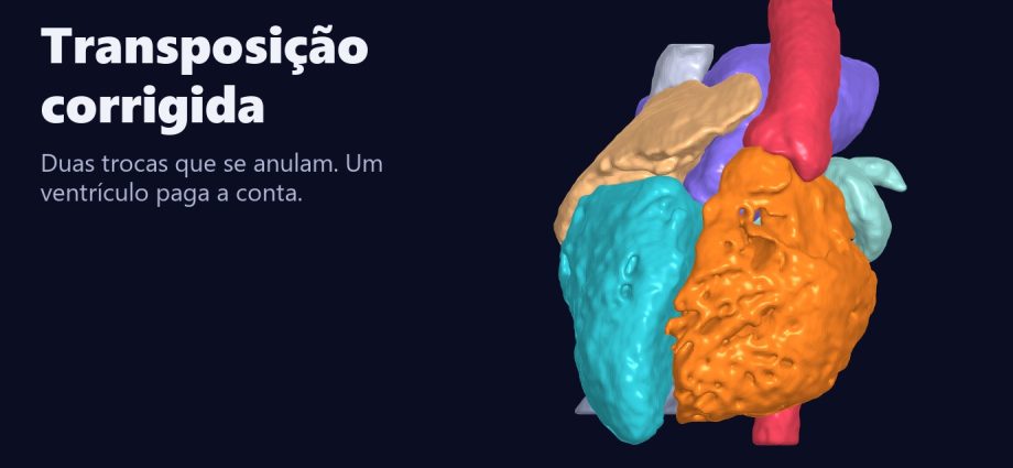
Transposição corrigida em 3D: duas trocas que se anulam e o ventrículo que paga a conta
Em um coração real de ressonância, o átrio direito entra no ventrículo esquerdo e o ventrículo direito sai na aorta. O sangue segue a rota certa, e por isso a doença ganhou o nome de corrigida. Mas a câmara que sustenta o corpo é um VD, com 323 cm² de superfície interna, e o nó AV está fora do lugar. O nome engana: aos 45 anos, 25% dos pacientes sem lesões associadas e 67% dos que as têm já estão em insuficiência cardíaca.
📅 Novembro de 2026
Navegue pelo Artigo
Da beira do leito ao coração real, e dele aos erros do caminho.
- 1. O diagnóstico que chega por uma bradicardia
- 2. Assista: Duas Trocas
- 3. Passeio 3D em nove passos
- 4. Duas trocas, uma rota certa
- 5. O coração real: o que a base mostrou
- 6. A aorta na frente e à esquerda
- 7. A armadilha: o nome
- 8. A fisiopatologia, passo a passo
- 9. O nó AV que mudou de endereço
- 10. O que fazer à beira do leito
- 11. Reparo fisiológico ou anatômico
- 12. Como foi feito e o que deu errado
- 13. Os números, sem filtro
- 14. O que é real e o que é calculado
- 15. Perguntas frequentes
- 16. Considerações Finais
- 17. Referências
1. O diagnóstico que chega por uma bradicardia
Um adolescente sem cianose chega ao pronto-socorro com síncope e frequência de 38. O ECG mostra bloqueio AV total e ondas Q nas precordiais direitas, que alguém lê como infarto inferior antigo [1]. Ninguém nunca tinha falado em cardiopatia, muito menos em transposição corrigida. (A cena é hipotética, mas cada peça dela está descrita na literatura.)
A transposição congenitamente corrigida é rara, cerca de 1 em 33.000 nascidos vivos [1], e pode ficar calada por décadas. Quando há lesões associadas ela aparece cedo. Quando não há, aparece tarde, às vezes como bloqueio, às vezes como falência do ventrículo sistêmico.
Para quem trabalha em UTI pediátrica, o risco é ler o ecocardiograma pela posição das câmaras e tratar um VD como se fosse VE.
2. Assista: Duas Trocas
Cerca de 100 segundos, em duas vozes. Duas setas de luz ligam cada átrio ao ventrículo trocado, o plano desce até a ressonância e os dois ventrículos se separam na mesma escala.
3. Passeio 3D em nove passos
Cada passo acende só o que importa nos dois corações reais, lado a lado e na mesma escala: um normal, de 17 anos, e a transposição corrigida, de 29. A bússola mostra sempre os lados do paciente, e o último passo é um autoteste.
Entenda em nove passos.
Siga os passos: cada um acende só o que importa nos dois corações reais, lado a lado. Depois gire, amplie e faça o teste.
O modelo só é baixado quando você abre.
4. Duas trocas, uma rota certa
O nome técnico é dupla discordância. O átrio direito se conecta ao ventrículo esquerdo morfológico, que ejeta na pulmonar. O átrio esquerdo se conecta ao ventrículo direito morfológico, que ejeta na aorta [1].
Na D-TGA há uma troca só, e os circuitos ficam em paralelo: sem mistura, o recém-nascido não sobrevive. Aqui há duas. Uma anula a outra, e os circuitos voltam a ficar em série.
Como ler direita e esquerda neste artigo
VD, VE, AD e AE são nomes pela morfologia da câmara, não pela posição: o VE deste coração fica do lado direito do paciente em relação ao VD. Quando o texto fala de lado, é sempre o lado do paciente. Nas imagens de frente e no corte, a direita do paciente aparece à esquerda da imagem, como em uma radiografia. “Aorta à esquerda” significa à esquerda da pulmonar.

Um exemplo concreto: o trem nos trilhos trocados
Dois desvios errados na mesma linha levam o trem à estação certa. O passageiro não percebe nada. Quem paga é a locomotiva de manobra, que agora puxa o trem de longa distância.
5. O coração real: o que a base mostrou
Usei o HVSMR-2.0, com 60 ressonâncias de cardiopatias congênitas segmentadas à mão por especialistas [8]. O caso escolhido é o de um adulto de 29 anos, com CIV e dextrocardia nos metadados. Não sei mais nada sobre essa pessoa, e não tentei saber.
Em vez de confiar na etiqueta, medi quantos voxels de cada câmara encostam na vizinha. Nesse coração, AD com VD dá zero. AE com VE dá zero. Toda a conexão atrioventricular passa pelo caminho cruzado.


No corte, o VE, do lado direito do paciente em relação ao VD (à esquerda na imagem), tem contorno liso. O VD tem trabéculas grossas, exatamente o critério morfológico que define a câmara [1].
| Contato (voxels) | Normal, 4 anos | D-TGA, 2 anos | Corrigida, 29 anos |
|---|---|---|---|
| AD com VD | 571 | 954 | 0 |
| AD com VE | 0 | 0 | 1.605 |
| AE com VE | 602 | 1.138 | 0 |
| AE com VD | 0 | 0 | 874 |
| VE com aorta | 244 | 0 | 0 |
| VD com aorta | 0 | 815 | 609 |
| VE com pulmonar | 0 | 67 | 491 |
| VD com pulmonar | 383 | 0 | 48 (ver seção 12) |
Os voxels têm tamanhos diferentes em cada exame, então o que vale nesta tabela é zero contra não zero. Contato de lúmen mostra vizinhança e não prova fluxo.
6. A aorta na frente e à esquerda
Vista de cima, a aorta deste coração sai 19,9 mm à frente da pulmonar e 26,7 mm à esquerda dela. A descrição clássica é essa mesma, anterior e à esquerda, embora nem todo caso a siga [1]. Por isso o nome L-TGA é impreciso: há corrigida com a aorta à direita.

As duas artérias sobem paralelas, sem a espiral do coração normal, o que deixa a borda superior da silhueta retificada na radiografia [1].
7. A armadilha: o nome
A palavra corrigida descreve a rota do sangue. Ela não diz nada sobre quem faz o trabalho. Uma revisão de 2023 foi direta no título: “not correct at all” [5].
Separei os dois ventrículos do mesmo coração e pus na mesma escala. O molde do VD tem 143 mL e 323 cm². O do VE, 102 mL e 169 cm². Com 1,4 vez o volume, o VD tem 1,9 vez a superfície, porque a parede é trabeculada.

Esse é um VD que continua VD depois de 29 anos contra a pressão sistêmica. Falta a ele a arquitetura helicoidal e a torção do VE [1].
Tradutor: o que dizem os números de longo prazo
Em 182 pacientes de 19 instituições, aos 45 anos 67% dos que tinham lesões associadas estavam em insuficiência cardíaca, contra 25% dos que não tinham [3]. Em 558 adultos seguidos por mediana de 8,7 anos, houve 15,4 eventos (assistência mecânica, transplante ou morte) por 1.000 pessoas-ano. Com disfunção de VD e insuficiência tricúspide moderadas ou graves foram 31 eventos em 110 pacientes. Com ambas leves ou ausentes, 13 em 181 [4]. São coortes retrospectivas.
Traduzindo para o chão da UTI: a tricúspide aqui é a valva sistêmica. Insuficiência que parece modesta num laudo pede a mesma atenção que uma insuficiência mitral num coração comum.
8. A fisiopatologia, passo a passo
Tudo começa no embrião. O tubo cardíaco costuma dobrar para a direita. Aqui ele dobra para a esquerda, e o VD vai parar à esquerda do VE [1]. É por isso que o AE encontra o VD. Ninguém sabe ainda por que esse desvio costuma vir junto com a troca das grandes artérias [1].
Sem outra lesão, a hemodinâmica pode ser normal [1]. O sangue percorre os dois circuitos em série, não há cianose, e a criança pode crescer sem sintomas. O problema fica escondido na carga de trabalho do ventrículo.
Por que o VD cansa
O VE é feito para pressão alta: suas fibras em hélice torcem a cavidade a cada sístole. O VD nasceu para empurrar sangue a um pulmão de baixa pressão, e não tem essa hélice nem essa torção [1]. Na transposição corrigida ele enfrenta a pressão sistêmica todos os dias, a vida inteira.
A resposta é hipertrofia e aumento da cavidade, e com os anos a função cai [1]. No esforço, esse VD não aumenta o trabalho como um VE normal, e a perfusão do miocárdio pode não acompanhar a demanda [1].
Um exemplo concreto: o ventilador de transporte
Ele ventila, e ventila bem. Mas ninguém o deixaria no leito por vinte anos. O VD sistêmico é esse equipamento: funciona, só que foi projetado para outra tarefa.
O ciclo da tricúspide
- Aqui a valva do lado sistêmico é a tricúspide, e ela tem alguma anomalia em até nove de cada dez pacientes [1].
- Costuma continuar competente na primeira década e passa a vazar entre a segunda e a quinta [1].
- Cada sístole devolve parte do sangue ao AE. Esse volume volta ao VD no batimento seguinte, e o VD passa a trabalhar com pressão alta e volume a mais [1].
- A pressão no AE sobe, e ela é transmitida para trás, até o pulmão.
- Insuficiência tricúspide e disfunção do VD andam juntas [3]. Na beira do leito, costuma ser difícil dizer qual veio primeiro.
E o VE, o que faz?
Ele trabalha contra a pressão pulmonar, baixa. Isso importa na hora de pensar em devolver o corpo a ele: em uma série de double switch, 12 de 46 pacientes precisaram de bandagem da pulmonar antes da cirurgia [9].
As lesões associadas mudam tudo
A transposição corrigida raramente vem sozinha. A CIV aparece em até três quintos dos pacientes, e a obstrução da saída do VE em dois quintos [1]. O que a criança apresenta depende dessa combinação.
| Combinação | O que acontece | Quando aparece |
|---|---|---|
| Sem lesão relevante | Rota certa, sem cianose; o VD pode falhar com os anos | Tarde [1] |
| CIV com repercussão | Insuficiência cardíaca | Cedo [1] |
| CIV e estenose pulmonar | O sangue azul encontra a saída do VE obstruída, cruza a CIV para o VD e vai à aorta: cianose | Cedo [1] |
| Tricúspide ebsteinoide | Insuficiência da valva sistêmica | Pode estar presente ao nascer [1] |
| Bloqueio AV | Bradicardia | Ao nascer ou ao longo da vida [1,2] |
Traduzindo para o chão da UTI: o que sobe a pós-carga sistêmica cai sobre o VD e sobre a tricúspide. Antes de subir o vasoconstritor nesse paciente, olhe a valva. É raciocínio fisiológico, não resultado de ensaio.
9. O nó AV que mudou de endereço
A malformação desloca o septo atrial em relação ao ventricular, e o feixe não consegue nascer do nó habitual. O nó fica anterior, sob a abertura do apêndice atrial direito, e o feixe longo desce pela face anterior da via de saída subpulmonar [1].
Em 107 pacientes, 22% tinham bloqueio AV total, com risco de cerca de 2% ao ano e septo intacto como fator de risco [2]. Cerca de um décimo já nasce com bloqueio [1].
O desvio do feixe explica também o ECG: a ativação septal se inverte, com Q nas precordiais direitas e sem Q nas esquerdas [1]. E explica por que fechar uma CIV aqui é outro procedimento, porque o feixe fica vulnerável durante o reparo [1].
10. O que fazer à beira do leito
- ☐ Nomeie cada ventrículo pela morfologia no prontuário e na passagem de plantão. “Ventrículo sistêmico (VD)” evita erro às três da manhã.
- ☐ Tenha um ECG de base com PR anotado. PR que alonga ao longo do tempo antecede o bloqueio total [1].
- ☐ No pós-operatório de CIV ou de via subpulmonar, espere bloqueio: até um quarto dos pacientes pode desenvolvê-lo na cirurgia [1]. Testar os fios de marca-passo antes de sair do centro cirúrgico é conduta prática, não dado de estudo.
- ☐ Cateter pela via subpulmonar pode causar bloqueio transitório [1].
- ☐ Marca-passo definitivo não é neutro: estimular o VE subpulmonar pode piorar o VD sistêmico. Uma série pediátrica de 7 pacientes com estimulação do sistema de condução preservou a função do VD em um ano [7], o que ainda é evidência fraca.
- ☐ Peça a avaliação da tricúspide em todo eco, e compare com o anterior.
11. Reparo fisiológico ou anatômico
O reparo fisiológico trata as lesões associadas e deixa o VD como sistêmico. O anatômico, o double switch, troca as vias atrial e arterial (ou usa Rastelli) para devolver o corpo ao VE [1].
Em 90 pacientes de uma instituição, a sobrevida em 20 anos foi semelhante, 75,7% com atrial mais Rastelli e 83,3% com atrial mais arterial, e a segunda opção teve menos reoperação e arritmia [6]. A revisão de 2023 aponta tendência a melhor sobrevida no reparo anatômico, com morbidade relevante nas duas estratégias [5], e o paciente sem lesão associada pode ir bem sem cirurgia por muito tempo. Não encontrei ensaio randomizado nas fontes que revisei, e a decisão segue dependendo de centro e de anatomia.
12. Como foi feito e o que deu errado
As segmentações foram reorientadas para o sistema anatômico, convertidas em malha e renderizadas em projeção ortográfica, para a escala em milímetros ser exata. Os contatos foram contados por dilatação de um voxel, e a posição das raízes é o centro do plano de contato entre ventrículo e artéria. As mesmas medidas reproduziram os 13,5 e 17,6 mm publicados no episódio da D-TGA.
A etiqueta não bastava. Os metadados clínicos da base marcam três pacientes como L-TGA. Medi os três. No de 12 anos, os dois átrios entram no VE: é dupla via de entrada, não dupla discordância. Outro tem átrio comum. Só um mostrou as duas trocas.
Quarenta e oito voxels sem explicação. O VD encosta 48 voxels na pulmonar, a 10,2 mm do plano da valva pulmonar. Pode ser a vizinhança da CIV subpulmonar, que é o tipo habitual aqui [1], ou só parede fina. O sinal da RM não resolveu, e deixei o número na tabela.
Casos deixados de fora. Dois pacientes com padrão sugestivo de cirurgia anatômica não têm metadados que fechem a história. Prefiro não contar o que não consigo conferir.
Três rótulos no lugar errado. No primeiro render do filme, os rótulos apontavam para o centro de cada câmara. Em 3D esse ponto pode estar atrás de outra estrutura: o AE caía sobre o VD, o AD sobre o VE e a pulmonar sobre o AD. Refiz os rótulos pela superfície visível e auditei cada um, quadro a quadro, contra o mapa de identidade da imagem.
A primeira figura dos ventrículos saiu cortada e descentrada, e um rótulo cobriu o átrio direito. Consertei os dois. Meu primeiro título falava em dois erros que se anulam. Troquei por trocas, porque é o coração de alguém.
13. Os números, sem filtro
| Medida | Valor | Origem |
|---|---|---|
| Aorta em relação à pulmonar | 19,9 mm à frente, 26,7 mm à esquerda | medida, 1 coração |
| Molde do VD | 143 mL, 323 cm² | medida |
| Molde do VE | 102 mL, 169 cm² | medida |
| Irregularidade VD/VE (área ÷ volume2/3) | 1,54 (normal 1,36; D-TGA 1,65) | medida, 3 corações |
| Incidência | cerca de 1 em 33.000 nascidos vivos | revisão [1] |
| Bloqueio AV total | 22% de 107; cerca de 2% ao ano | coorte [2] |
| Insuficiência cardíaca aos 45 anos | 67% com lesões; 25% sem | 182 pacientes [3] |
| Desfecho composto | 15,4 por 1.000 pessoas-ano | 558 adultos [4] |
| Sobrevida em 20 anos, double switch | 75,7% e 83,3% | 90 pacientes [6] |
14. O que é real e o que é calculado
⚠ Limites
Real: a ressonância, a forma das câmaras e a segmentação manual da base. As máscaras são do lúmen, sem miocárdio.
Calculado: contatos, volumes, áreas e distâncias. Área depende de resolução e de suavização, então a razão entre ventrículos do mesmo exame é mais confiável que o valor absoluto.
Esquema: o diagrama de circuitos. Nenhum fluxo foi medido, nenhuma função foi avaliada, e a fase cardíaca não consta nos metadados.
Um coração não representa a doença. Material educacional, sem uso diagnóstico.
15. Perguntas frequentes
Transposição corrigida dá cianose?
Sozinha, não. A cianose vem de CIV com estenose pulmonar [1].
É a mesma coisa que L-TGA?
Costuma ser usada assim, mas o termo é impreciso: há casos com a aorta à direita, e com átrios em imagem em espelho [1].
Todo paciente precisa de cirurgia?
O diagnóstico sozinho não indica operação [1]. A decisão depende das lesões associadas e do risco de falência do VD.
Por que o paciente pode descobrir só na vida adulta?
Porque a rota do sangue está certa. O primeiro sinal pode ser bloqueio ou insuficiência cardíaca [1].
Quer ensinar anatomia assim?
A Metodologia AIMED organiza o uso de IA em medicina, com revisão clínica em cada etapa.
16. Considerações Finais
Todo mundo decora “dupla discordância”. Poucos olham o que isso custa: 323 cm² de trabécula empurrando contra a pressão sistêmica, com o nó AV em outro endereço.
💡 Connecting the Dots. O dado que se cita é o da rota certa. O que pouca gente traduz é que a rota certa atrasa o diagnóstico e dá ao ventrículo errado décadas de trabalho. Medi isso em um coração, com um método que qualquer colega pode repetir sobre dados abertos, e dois dos três casos marcados como L-TGA não eram o que eu procurava. Conferir antes de ensinar custa uma tarde.
Este coração só pôde ser estudado porque o exame de alguém está em uma base pública.
Referências
- Wallis GA, Debich-Spicer D, Anderson RH. Congenitally corrected transposition. Orphanet J Rare Dis. 2011;6:22. PMID: 21569592. DOI: 10.1186/1750-1172-6-22
- Huhta JC, Maloney JD, Ritter DG, Ilstrup DM, Feldt RH. Complete atrioventricular block in patients with atrioventricular discordance. Circulation. 1983;67(6):1374-7. PMID: 6851033. DOI: 10.1161/01.cir.67.6.1374
- Graham TP, Bernard YD, Mellen BG, et al. Long-term outcome in congenitally corrected transposition of the great arteries: a multi-institutional study. J Am Coll Cardiol. 2000;36(1):255-61. PMID: 10898443. DOI: 10.1016/s0735-1097(00)00682-3
- van Dissel AC, Opotowsky AR, Burchill LJ, et al. End-stage heart failure in congenitally corrected transposition of the great arteries: a multicentre study. Eur Heart J. 2023;44(34):3278-91. PMID: 37592821. DOI: 10.1093/eurheartj/ehad511
- DeWeert KJ, Lancaster T, Dorfman AL. Congenitally corrected transposition: not correct at all. Curr Opin Cardiol. 2023;38(4):358-63. PMID: 37016955. DOI: 10.1097/HCO.0000000000001052
- Hiramatsu T, Matsumura G, Konuma T, et al. Long-term prognosis of double-switch operation for congenitally corrected transposition of the great arteries. Eur J Cardiothorac Surg. 2012;42(6):1004-8. PMID: 22551964. DOI: 10.1093/ejcts/ezs118
- Silvetti MS, Favoccia C, Saputo FA, et al. Three-dimensional-mapping-guided permanent conduction system pacing in paediatric patients with congenitally corrected transposition of the great arteries. Europace. 2023;25(4):1482-90. PMID: 36794445. DOI: 10.1093/europace/euad026
- Pace DF, Contreras HTM, Romanowicz J, et al. HVSMR-2.0. Sci Data. 2024;11:721. DOI: 10.1038/s41597-024-03469-9
- Duncan BW, Mee RB, Mesia CI, et al. Results of the double switch operation for congenitally corrected transposition of the great arteries. Eur J Cardiothorac Surg. 2003;24(1):11-9. PMID: 12853040. DOI: 10.1016/s1010-7940(03)00189-1
Música do filme: ‘Katabasis_II’ by Scott Buckley – released under CC-BY 4.0. www.scottbuckley.com.au
Corrected transposition in 3D: two swaps cancel out, and one ventricle pays the price
In a real MRI heart, the right atrium enters the left ventricle and the right ventricle exits into the aorta. Blood follows the right route, which is why the disease was named corrected. But the chamber supporting the body is an RV, with 323 cm² of inner surface, and the AV node is misplaced. The name misleads: by age 45, 25% of patients without associated lesions and 67% of those with them already have heart failure.
📅 November 2026
Article Guide
From the bedside to a real heart, and from there to the mistakes along the way.
- 1. The diagnosis that arrives as bradycardia
- 2. Watch: Two Swaps
- 3. Nine-step 3D tour
- 4. Two swaps, one right route
- 5. The real heart: what the dataset showed
- 6. The aorta in front and to the left
- 7. The trap: the name
- 8. The pathophysiology, step by step
- 9. The AV node that changed address
- 10. What to do at the bedside
- 11. Physiologic or anatomic repair
- 12. How it was done and what went wrong
- 13. The numbers, unfiltered
- 14. What is real and what is computed
- 15. Frequently asked questions
- 16. Final Thoughts
- 17. References
1. The diagnosis that arrives as bradycardia
A teenager with no cyanosis reaches the emergency room after syncope, heart rate 38. The ECG shows complete AV block and Q waves in the right precordial leads, which someone reads as an old inferior infarction [1]. Nobody had ever mentioned heart disease, let alone corrected transposition. (The scene is hypothetical, but each piece of it is described in the literature.)
Congenitally corrected transposition is rare, about 1 in 33,000 live births [1], and it can stay silent for decades. With associated lesions it shows up early. Without them it shows up late, sometimes as block, sometimes as failure of the systemic ventricle.
For anyone working in a pediatric ICU, the risk is reading the echo by chamber position and treating an RV as if it were an LV.
2. Watch: Two Swaps
About 110 seconds, in two voices. Two arrows of light link each atrium to its swapped ventricle, the plane descends to the MRI, and the two ventricles separate at the same scale.
3. Nine-step 3D tour
Each step lights only what matters in the two real hearts, side by side and at the same scale: a normal heart, age 17, and corrected transposition, age 29. The compass always shows the patient sides, and the last step is a self-test.
Understand it in nine steps.
Follow the steps: each one lights only what matters in the two real hearts, side by side. Then rotate, zoom and take the test.
The model downloads only when you open it.
4. Two swaps, one right route
The technical name is double discordance. The right atrium connects to the morphological left ventricle, which ejects into the pulmonary artery. The left atrium connects to the morphological right ventricle, which ejects into the aorta [1].
In D-TGA there is a single swap, and the circuits run in parallel: without mixing, the newborn does not survive. Here there are two. One cancels the other, and the circuits return to series.
How to read right and left in this article
RV, LV, RA and LA name the chamber by morphology, not by position: the LV of this heart lies on the patient’s right relative to the RV. When the text speaks of a side, it is always the patient’s side. In frontal images and on the slice, the patient’s right appears on image left, as on a chest film. “Aorta to the left” means to the left of the pulmonary artery.

A concrete example: the train on swapped tracks
Two wrong switches on the same line take the train to the right station. The passenger notices nothing. The shunting engine pays, because it now hauls the long-distance train.
5. The real heart: what the dataset showed
I used HVSMR-2.0, with 60 MRI scans of congenital heart disease segmented by hand by experts [8]. The chosen case is a 29-year-old adult, with VSD and dextrocardia in the metadata. I know nothing else about this person, and I did not try to find out.
Instead of trusting the label, I measured how many voxels of each chamber touch its neighbor. In this heart, RA with RV gives zero. LA with LV gives zero. All atrioventricular connection runs through the crossed route.


On the slice, the LV, on the patient’s right relative to the RV (image left), has a smooth outline. The RV has coarse trabeculations, exactly the morphological criterion that defines the chamber [1].
| Contact (voxels) | Normal, age 4 | D-TGA, age 2 | Corrected, age 29 |
|---|---|---|---|
| RA with RV | 571 | 954 | 0 |
| RA with LV | 0 | 0 | 1,605 |
| LA with LV | 602 | 1,138 | 0 |
| LA with RV | 0 | 0 | 874 |
| LV with aorta | 244 | 0 | 0 |
| RV with aorta | 0 | 815 | 609 |
| LV with pulmonary | 0 | 67 | 491 |
| RV with pulmonary | 383 | 0 | 48 (see section 12) |
Voxel size differs between scans, so what counts in this table is zero against non-zero. Lumen contact shows adjacency and does not prove flow.
6. The aorta in front and to the left
Seen from above, the aorta of this heart leaves 19.9 mm in front of the pulmonary artery and 26.7 mm to its left. That matches the classic description, anterior and leftward, although not every case follows it [1]. This is why the name L-TGA is imprecise: corrected transposition exists with a right-sided aorta.

The two arteries rise in parallel, without the spiral of the normal heart, which leaves the upper border of the silhouette straightened on the chest film [1].
7. The trap: the name
The word corrected describes the route of the blood. It says nothing about who does the work. A 2023 review was direct in its title: “not correct at all” [5].
I separated the two ventricles of the same heart and put them at the same scale. The RV cast has 143 mL and 323 cm². The LV cast, 102 mL and 169 cm². With 1.4 times the volume, the RV has 1.9 times the surface, because its wall is trabeculated.

This is an RV that is still an RV after 29 years against systemic pressure. It lacks the helical architecture and the torsion of the LV [1].
Translator: what the long-term numbers say
In 182 patients from 19 institutions, by age 45, 67% of those with associated lesions had heart failure, against 25% of those without [3]. In 558 adults followed for a median of 8.7 years, there were 15.4 events (mechanical support, transplant or death) per 1,000 person-years. With moderate or severe RV dysfunction and tricuspid regurgitation there were 31 events in 110 patients. With both mild or absent, 13 in 181 [4]. These are retrospective cohorts.
Translating to the ICU floor: the tricuspid valve here is the systemic valve. Regurgitation that looks modest on a report deserves the attention you would give mitral regurgitation in an ordinary heart.
8. The pathophysiology, step by step
It all begins in the embryo. The heart tube usually bends to the right. Here it bends to the left, and the RV ends up to the left of the LV [1]. That is why the LA meets the RV. Nobody yet knows why this deviation so often comes with the swap of the great arteries [1].
With no other lesion, hemodynamics can be normal [1]. Blood runs through the two circuits in series, there is no cyanosis, and the child may grow up without symptoms. The problem stays hidden in the ventricle’s workload.
Why the RV tires
The LV is built for high pressure: its helical fibers twist the cavity with each systole. The RV was born to push blood into a low-pressure lung, and it has neither that helix nor that torsion [1]. In corrected transposition it faces systemic pressure every day, for life.
The response is hypertrophy and cavity enlargement, and over the years function falls [1]. During exercise, this RV does not increase its work the way a normal LV does, and myocardial perfusion may not keep up with demand [1].
A concrete example: the transport ventilator
It ventilates, and it ventilates well. But nobody would leave it at the bedside for twenty years. The systemic RV is that device: it works, but it was designed for another job.
The tricuspid cycle
- Here the valve on the systemic side is the tricuspid, and it has some anomaly in up to nine of every ten patients [1].
- It usually stays competent in the first decade and starts to leak between the second and fifth [1].
- Each systole sends part of the blood back to the LA. That volume returns to the RV on the next beat, so the RV works with high pressure and extra volume [1].
- LA pressure rises, and it is transmitted backward to the lungs.
- Tricuspid regurgitation and RV dysfunction go together [3]. At the bedside, it is often hard to say which came first.
And what does the LV do?
It works against low pulmonary pressure. That matters when thinking about giving the body back to it: in one double switch series, 12 of 46 patients needed pulmonary artery banding before surgery [9].
Associated lesions change everything
Corrected transposition rarely comes alone. A VSD appears in up to three fifths of patients, and LV outflow obstruction in two fifths [1]. What the child presents with depends on that combination.
| Combination | What happens | When it appears |
|---|---|---|
| No relevant lesion | Right route, no cyanosis; the RV may fail over the years | Late [1] |
| Hemodynamically significant VSD | Heart failure | Early [1] |
| VSD and pulmonary stenosis | Blue blood meets an obstructed LV outlet, crosses the VSD to the RV and reaches the aorta: cyanosis | Early [1] |
| Ebstein-like tricuspid | Systemic valve regurgitation | May be present at birth [1] |
| AV block | Bradycardia | At birth or through life [1,2] |
Translating to the ICU floor: whatever raises systemic afterload falls on the RV and the tricuspid valve. Before turning up the vasoconstrictor in this patient, look at the valve. This is physiological reasoning, not a trial result.
9. The AV node that changed address
The malformation shifts the atrial septum relative to the ventricular septum, and the bundle cannot arise from the usual node. The node sits anteriorly, beneath the opening of the right atrial appendage, and the long bundle runs down the anterior face of the subpulmonary outflow tract [1].
In 107 patients, 22% had complete AV block, with a risk of about 2% per year and an intact septum as a risk factor [2]. About one tenth are born with block [1].
The bundle’s detour also explains the ECG: septal activation reverses, with Q waves in the right precordial leads and none in the left [1]. And it explains why closing a VSD here is a different procedure, because the bundle is vulnerable during the repair [1].
10. What to do at the bedside
- ☐ Name each ventricle by morphology in the chart and at handover. “Systemic ventricle (RV)” prevents errors at three in the morning.
- ☐ Keep a baseline ECG with the PR written down. A PR that lengthens over time precedes complete block [1].
- ☐ After VSD or subpulmonary outflow surgery, expect block: up to a quarter of patients may develop it during surgery [1]. Testing pacing wires before leaving the operating room is practical care, not a study finding.
- ☐ A catheter through the subpulmonary route can cause transient block [1].
- ☐ A permanent pacemaker is not neutral: pacing the subpulmonary LV can worsen the systemic RV. A pediatric series of 7 patients with conduction system pacing preserved RV function at one year [7], which is still weak evidence.
- ☐ Ask for a tricuspid assessment on every echo, and compare it with the previous one.
11. Physiologic or anatomic repair
Physiologic repair treats the associated lesions and leaves the RV as systemic. Anatomic repair, the double switch, redirects the atrial and arterial routes (or uses a Rastelli) to give the body back to the LV [1].
In 90 patients from one institution, 20-year survival was similar, 75.7% with atrial plus Rastelli and 83.3% with atrial plus arterial, and the second option had less reoperation and arrhythmia [6]. The 2023 review points to a trend toward better survival with anatomic repair, with relevant morbidity in both strategies [5], and a patient with no associated lesion may do well without surgery for a long time. I found no randomized trial in the sources I reviewed, and the decision still depends on center and anatomy.
12. How it was done and what went wrong
The segmentations were reoriented to the anatomical system, converted to meshes and rendered in orthographic projection, so the millimeter scale is exact. Contacts were counted by one-voxel dilation, and root position is the center of the contact plane between ventricle and artery. The same measurements reproduced the 13.5 and 17.6 mm published in the D-TGA episode.
The label was not enough. The dataset’s clinical metadata mark three patients as L-TGA. I measured all three. In the 12-year-old, both atria enter the LV: that is double inlet, not double discordance. Another has a common atrium. Only one showed both swaps.
Forty-eight unexplained voxels. The RV touches the pulmonary artery over 48 voxels, 10.2 mm from the pulmonary valve plane. It may be the neighborhood of a subpulmonary VSD, the usual type here [1], or just a thin wall. The MRI signal did not settle it, and I left the number in the table.
Cases left out. Two patients with a pattern suggesting anatomic surgery have metadata that do not close the story. I would rather not tell what I cannot check.
Three labels in the wrong place. In the first render of the film, labels pointed to the center of each chamber. In 3D that point can sit behind another structure: the LA landed on the RV, the RA on the LV and the pulmonary artery on the RA. I rebuilt the labels on the visible surface and audited each one, frame by frame, against the image identity map.
The first ventricle figure came out cropped and off-center, and a label covered the right atrium. I fixed both. My first title spoke of two errors that cancel out. I changed it to swaps, because this is someone’s heart.
13. The numbers, unfiltered
| Measure | Value | Source |
|---|---|---|
| Aorta relative to pulmonary artery | 19.9 mm in front, 26.7 mm to the left | measured, 1 heart |
| RV cast | 143 mL, 323 cm² | measured |
| LV cast | 102 mL, 169 cm² | measured |
| RV/LV irregularity (area ÷ volume2/3) | 1.54 (normal 1.36; D-TGA 1.65) | measured, 3 hearts |
| Incidence | about 1 in 33,000 live births | review [1] |
| Complete AV block | 22% of 107; about 2% per year | cohort [2] |
| Heart failure by age 45 | 67% with lesions; 25% without | 182 patients [3] |
| Composite outcome | 15.4 per 1,000 person-years | 558 adults [4] |
| 20-year survival, double switch | 75.7% and 83.3% | 90 patients [6] |
14. What is real and what is computed
⚠ Limits
Real: the MRI, the shape of the chambers and the dataset’s manual segmentation. Masks are of the lumen, without myocardium.
Computed: contacts, volumes, areas and distances. Area depends on resolution and smoothing, so the ratio between ventricles of the same scan is more reliable than the absolute value.
Diagram: the circuit figure. No flow was measured, no function was assessed, and cardiac phase is not in the metadata.
One heart does not represent the disease. Educational material, not for diagnostic use.
15. Frequently asked questions
Does corrected transposition cause cyanosis?
Not on its own. Cyanosis comes from a VSD with pulmonary stenosis [1].
Is it the same as L-TGA?
The terms are often used that way, but L-TGA is imprecise: there are cases with a right-sided aorta and with mirror-image atria [1].
Does every patient need surgery?
The diagnosis alone does not indicate an operation [1]. The decision depends on associated lesions and the risk of RV failure.
Why might a patient only find out as an adult?
Because the route of the blood is right. The first sign may be block or heart failure [1].
Want to teach anatomy this way?
The AIMED Methodology organizes the use of AI in medicine, with clinical review at every step.
16. Final Thoughts
Everyone memorizes “double discordance”. Few look at what it costs: 323 cm² of trabeculations pushing against systemic pressure, with the AV node at another address.
💡 Connecting the Dots. The fact people quote is the right route. What few translate is that the right route delays diagnosis and gives the wrong ventricle decades of work. I measured this in one heart, with a method any colleague can repeat on open data, and two of the three cases marked L-TGA were not what I was looking for. Checking before teaching costs an afternoon.
This heart could only be studied because someone’s scan is in a public dataset.
References
- Wallis GA, Debich-Spicer D, Anderson RH. Congenitally corrected transposition. Orphanet J Rare Dis. 2011;6:22. PMID: 21569592. DOI: 10.1186/1750-1172-6-22
- Huhta JC, Maloney JD, Ritter DG, Ilstrup DM, Feldt RH. Complete atrioventricular block in patients with atrioventricular discordance. Circulation. 1983;67(6):1374-7. PMID: 6851033. DOI: 10.1161/01.cir.67.6.1374
- Graham TP, Bernard YD, Mellen BG, et al. Long-term outcome in congenitally corrected transposition of the great arteries: a multi-institutional study. J Am Coll Cardiol. 2000;36(1):255-61. PMID: 10898443. DOI: 10.1016/s0735-1097(00)00682-3
- van Dissel AC, Opotowsky AR, Burchill LJ, et al. End-stage heart failure in congenitally corrected transposition of the great arteries: a multicentre study. Eur Heart J. 2023;44(34):3278-91. PMID: 37592821. DOI: 10.1093/eurheartj/ehad511
- DeWeert KJ, Lancaster T, Dorfman AL. Congenitally corrected transposition: not correct at all. Curr Opin Cardiol. 2023;38(4):358-63. PMID: 37016955. DOI: 10.1097/HCO.0000000000001052
- Hiramatsu T, Matsumura G, Konuma T, et al. Long-term prognosis of double-switch operation for congenitally corrected transposition of the great arteries. Eur J Cardiothorac Surg. 2012;42(6):1004-8. PMID: 22551964. DOI: 10.1093/ejcts/ezs118
- Silvetti MS, Favoccia C, Saputo FA, et al. Three-dimensional-mapping-guided permanent conduction system pacing in paediatric patients with congenitally corrected transposition of the great arteries. Europace. 2023;25(4):1482-90. PMID: 36794445. DOI: 10.1093/europace/euad026
- Pace DF, Contreras HTM, Romanowicz J, et al. HVSMR-2.0. Sci Data. 2024;11:721. DOI: 10.1038/s41597-024-03469-9
- Duncan BW, Mee RB, Mesia CI, et al. Results of the double switch operation for congenitally corrected transposition of the great arteries. Eur J Cardiothorac Surg. 2003;24(1):11-9. PMID: 12853040. DOI: 10.1016/s1010-7940(03)00189-1
Música do filme: ‘Katabasis_II’ by Scott Buckley – released under CC-BY 4.0. www.scottbuckley.com.au
◆ Novidades
- Anatomia do olho em 3D: faça o teste do olhinho em um olho realNOVO
- Anatomia da cabeça em 3D: rosto, crânio e cérebro de uma só pessoaNOVO
- Fontan em 3D: o caminho até o pulmão que não passa pelo coração