
Por que apertar a artéria de um bebê? A bandagem da pulmonar num coração real
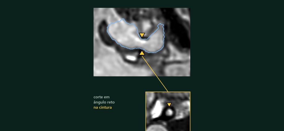
Na ressonância real de um bebê com dupla via de entrada do ventrículo esquerdo e bandagem da artéria pulmonar, o tronco mede 10,2 mm antes da faixa, 3,6 mm na faixa e 7,6 mm depois, cerca de metade do calibre vizinho. Esse número está no filme. Se a faixa está apertada o suficiente, a imagem não diz (isso fica com o Doppler), e nesse tipo de coração existe um risco descrito do outro lado, na saída para a aorta.
📅 Novembro de 2026
Navegue pelo Artigo
Da passagem de plantão às medidas, com o que ficou de fora delas.
- 1. Por que isso importa no plantão
- 2. Assista: A Faixa
- 3. Passeio 3D em oito passos
- 4. O que é a bandagem da artéria pulmonar
- 5. O coração real, em números
- 6. A cintura medida
- 7. O que o Doppler mostra após a bandagem
- 8. O risco do outro lado: a saída para a aorta
- 9. Traduzindo para o chão da UTI
- 10. Como foi feito e o que deu errado
- 11. O que é real e o que é calculado
- 12. Perguntas frequentes
- 13. Considerações Finais
- 14. Referências
1. Por que isso importa no plantão
Duas da manhã. Chega à UTI um lactente com coração de ventrículo único, saturação boa demais para a cardiopatia e respiração cansada. A passagem de plantão termina com uma frase curta: “fez bandagem da pulmonar hoje”.
A pergunta do residente é a do título do filme: por que apertar a artéria de um bebê? E a pergunta seguinte, a que define a noite, é outra: essa faixa está apertada o suficiente, ou demais?
Fui atrás de um coração real com essa faixa, num banco público de ressonâncias, para ver quanto ela de fato estreita a artéria. Medi o que a imagem permite medir. O resto, deixei escrito como limite.
2. Assista: A Faixa
Noventa segundos, ressonância real, narração em português. A medida só aparece depois da pausa do quiz: responda antes de continuar.
3. Passeio 3D em oito passos
Os dois corações, lado a lado e na mesma escala: um de referência, com 4 anos, e o do bebê com bandagem. Cada passo acende só o que importa; a cintura da faixa pulsa em amarelo. No fim, um teste de três toques.
Por que apertar a artéria de um bebê? Descubra em oito passos.
Dois corações reais, lado a lado e na mesma escala. Cada passo acende só o que importa; a cintura da faixa pulsa em amarelo. Depois gire, amplie e faça o teste.
O modelo só é baixado quando você abre.
4. O que é a bandagem da artéria pulmonar
É uma faixa colocada em volta do tronco da artéria pulmonar para estreitá-lo. Em corações com um ventrículo só e fluxo pulmonar sem limite, o pulmão pode receber sangue demais, sob pressão alta. Com o tempo, a resistência dos vasos pulmonares sobe. E o caminho que muitos desses corações seguem, Glenn e depois Fontan, só funciona com pulmão de baixa resistência [1].
A faixa compra esse tempo. Não é a única estratégia: em pacientes selecionados, há séries que preferiram ir direto ao Glenn, sem banda [2], e a própria bandagem tem taxa relevante de reajuste antes do segundo estágio [3]. A escolha é do time cirúrgico, caso a caso.
Tradutor de siglas, pela pergunta do plantão
DILV (dupla via de entrada do ventrículo esquerdo): os dois átrios esvaziam no mesmo ventrículo. Glenn: a veia cava superior passa a drenar direto na artéria pulmonar. Fontan: o retorno venoso inteiro chega ao pulmão sem passar por um ventrículo. Em todos, o pulmão precisa deixar o sangue passar quase sem resistência.

5. O coração real, em números
O caso é o pat55 do banco público HVSMR-2.0 [12]: um bebê, sem idade em meses informada, com o diagnóstico registrado de comunicação interatrial, transposição, dupla via de entrada do ventrículo esquerdo e bandagem da artéria pulmonar. A anatomia é real e anonimizada; a história clínica do filme é ilustrativa.
| Medida | Bebê com bandagem | Referência (4 anos) | Leitura |
|---|---|---|---|
| Contato dos átrios com o ventrículo grande (voxels) | esquerdo 297 · direito 305 | cada átrio no seu ventrículo | → dupla via de entrada |
| Contato dos átrios com a câmara menor (voxels) | 0 e 3 | não se aplica | → câmara sem via de entrada |
| Volume do lúmen do ventrículo grande · câmara menor | 24 mL · 3,4 mL | 28 mL · 30 mL | ↓ câmara menor |
| De onde sai a artéria pulmonar | ventrículo grande (303 voxels de contato) | ventrículo direito | → relação trocada |
| De onde sai a aorta | câmara menor (621 voxels de contato) | ventrículo esquerdo | → relação trocada |
Contato de superfícies mostra conexão anatômica entre as máscaras; não mede fluxo nem o tamanho de uma passagem.

6. A cintura medida
Um plano em ângulo reto com a artéria anda por dentro do tronco, milímetro a milímetro. Em cada posição, mede-se o maior círculo que cabe no lúmen: o diâmetro inscrito. O mesmo método vale para o coração de referência.
| Posição | Bebê com bandagem | Referência | Leitura |
|---|---|---|---|
| Antes da faixa | 10,2 mm | 16,6 mm | → calibre de partida |
| Na cintura | 3,6 mm | 14,3 mm (meio do tronco) | ↓ cintura |
| Depois da faixa | 7,6 mm | 14,8 mm | → reabre |
| Razão cintura ÷ calibre depois | 0,47 | 0,97 | ↓ cerca de metade |
A cintura do bebê ocupa uns 3 mm de comprimento. No corte em ângulo reto, a luz é pequena e redonda, cercada por um anel escuro compatível com a parede e a faixa. No coração de referência, o mesmo plano atravessa o tronco inteiro sem encontrar cintura.

Diâmetro inscrito, pela pergunta do plantão
“Quanto passa por ali?” O diâmetro inscrito é o tamanho da maior bola que cabe no lúmen naquele corte. Mede a luz por dentro; a parede, a faixa e a pressão ficam fora da conta.
Um exemplo concreto: “a mesma escala”
No filme, os dois cortes aparecem lado a lado. A primeira versão tinha um erro: cada corte estava numa escala própria, 22 mm de um lado e 35 mm do outro. A comparação só ficou honesta quando os dois passaram a ocupar a tela na mesma escala em milímetros.

7. O que o Doppler mostra após a bandagem
Na UTI, saber quanto o tronco estreitou ajuda pouco sozinho. O que se quer saber é se a faixa está apertada o suficiente, e isso depende de pressão. A ressonância anatômica mostra a forma e para por aí: pressão, gradiente e fluxo ficam de fora.
Quem estima a diferença de pressão através da faixa é o Doppler. O eco mede a velocidade máxima do jato e a converte em gradiente pela equação de Bernoulli modificada. Num estudo com 20 pacientes submetidos à bandagem, o gradiente pelo Doppler contínuo comparado ao do cateterismo simultâneo teve correlação de 0,98, com erro-padrão de 7 mmHg [9]. Em outra série, com 37 crianças investigadas por suspeita de estenose valvar pulmonar ou para avaliação de uma bandagem, o Doppler também mostrou boa concordância com o cateterismo [10].
⚠ Ressalva
O Doppler estima o gradiente instantâneo; por isso bate melhor com o máximo instantâneo do cateter do que com o “pico a pico” [9]. Quando as grandes artérias estão em posição anômala, alinhar o feixe com o jato exige várias janelas [11]. E nenhum desses dados existe para este bebê: a base pública traz só a ressonância.
Num estudo retrospectivo de 50 pacientes com ventrículo único, as pressões pulmonares e a relação entre pressão pulmonar e sistêmica depois da banda diferiram entre quem progrediu para o estágio seguinte e quem não progrediu [4]. O ajuste fino da faixa, portanto, se conversa em mmHg.
8. O risco do outro lado: a saída para a aorta
Neste coração, a aorta sai da câmara menor. O sangue do ventrículo grande precisa atravessar uma passagem até lá. A literatura descreve que, depois da bandagem, essa passagem pode estreitar, e a obstrução pode passar a afetar também a saída para o corpo [1, 6, 7].
Em cinco pacientes com esse tipo de conexão, estudados por eco transesofágico durante a cirurgia, a redução do volume do ventrículo alterou a geometria na hora, diminuiu a passagem e gerou um gradiente mediano de 60 mmHg através dela [5]. Por isso, a bandagem neonatal nesse cenário costuma ser reservada a passagens grandes e livres, com vigilância ecocardiográfica próxima [6]. Numa coorte retrospectiva com risco de obstrução da saída sistêmica, a operação de Damus-Kaye-Stansel feita sem banda prévia teve sobrevida em dez anos maior do que a feita depois da banda [8].
Neste caso
A passagem entre o ventrículo grande e a câmara menor não foi medida. O filme mostra o risco como possibilidade descrita na literatura, em esquema, e mostra separadamente o coração real com a passagem tracejada, sem número.
9. Traduzindo para o chão da UTI
A medida do lúmen localiza e descreve a cintura. À beira do leito, o gradiente estimado pelo Doppler é interpretado junto com a saturação, a perfusão e o fluxo na saída para a aorta.
- ☐ Saturação e sinais de hiperfluxo ou de faixa apertada demais, na tendência das horas, não num valor isolado.
- ☐ Gradiente através da faixa pelo Doppler, sempre com a janela e o alinhamento descritos no laudo.
- ☐ Gradiente na saída para a aorta, também pelo eco, porque o risco descrito está do outro lado do coração.
- ☐ Lembrar que reajuste da faixa antes do segundo estágio não é raro [3].
Material educativo. As decisões são sempre do médico assistente e da equipe que conhece o paciente.
10. Como foi feito e o que deu errado
A triagem percorreu os 59 corações do banco. Entre os seis com bandagem registrada, cinco tinham a cintura entre 0,48 e 0,54 do calibre vizinho; nos cinco corações normais, a razão ficou entre 0,94 e 1,00.
Errei mais de uma vez até chegar a esses números. Conto os erros porque eles explicam as medidas:
- Dois milímetros fora. O primeiro método mediu a artéria em cascas a partir da raiz e marcou a cintura 2 mm antes do lugar certo. A revisão pediu cortes em ângulo reto ao vaso, e a cintura verdadeira apareceu um pouco adiante.
- Três números para a mesma cintura. 3,8 mm pelas cascas, 3,6 mm pela máscara no plano perpendicular e 2,7 mm pela borda do próprio sinal da ressonância. É a borda de uma luz com poucos pixels. O filme usa um método só, do começo ao fim.
- Um bebê quase descartado. Outro caso do banco parecia ter cintura fraca no perfil 3D. No plano perpendicular, o estreitamento existia, mas ficava colado à bifurcação.
- A voz e o texto. A narração foi gravada três vezes, até soar como uma professora que convida a raciocinar junto. O texto da anatomia foi reescrito em frases curtas que seguem a imagem.

11. O que é real e o que é calculado
| Elemento | Origem |
|---|---|
| Cortes da ressonância e superfícies dos corações | Real: HVSMR-2.0, segmentação dos autores da base [12] |
| Diâmetros e razões | Calculados sobre a segmentação, em planos perpendiculares |
| Contorno azul e tom da artéria pulmonar | Segmentação, identificada na tela |
| Fluxo para o pulmão, Doppler e passagem que estreita | Esquemas, marcados como tal |
| História clínica | Ilustrativa |
| Voz | Sintética (Gemini TTS); roteiro e revisão do Dr. Mbula |
Com voxels de 0,5 a 0,73 mm, a borda muda o número da cintura em cerca de 1 mm. Por isso o intervalo 2,7 a 3,8 mm aparece como estimativa conforme o método, e não como intervalo de confiança. A revisão por cardiologista pediátrico ou ecocardiografista é recomendada.
12. Perguntas frequentes
A faixa deste bebê está bem ajustada?
A ressonância não permite dizer. O ajuste depende da pressão, que se estima pelo Doppler ou se mede no cateterismo [9, 10].
Os 3,6 mm são o tamanho da faixa?
Não. São o diâmetro inscrito do lúmen no plano perpendicular. A faixa em si não aparece na segmentação.
Este bebê tem obstrução subaórtica?
Não sabemos. A passagem até a aorta não foi medida. O filme trata o risco como possibilidade descrita na literatura.
A IA fez o diagnóstico?
Não. O diagnóstico vem do registro da base, e a segmentação é dos autores. Os scripts medem o que a segmentação mostra, e a leitura foi revisada, com os erros expostos acima.
Quer ensinar anatomia assim?
A Metodologia AIMED organiza o uso de IA em medicina, com revisão clínica em cada etapa.
13. Considerações Finais
“Por que apertar a artéria de um bebê?” tem resposta clara: para proteger um pulmão que, um dia, vai precisar de baixa resistência. A resposta difícil é a seguinte: quanto apertar.
💡 Connecting the Dots. Todo mundo mostra a cintura, porque é a parte que se vê. Pouca gente diz em voz alta que a imagem anatômica só responde à geometria. “Cerca de metade do calibre vizinho” é um dado verdadeiro, e mesmo assim não decide nada à beira do leito. Quem orienta a conduta é o gradiente estimado pelo Doppler, lido junto com a avaliação da saída para a aorta. Eu fiz a parte da forma, com o cuidado de deixar escrito onde ela acaba; a pergunta da pressão continua com o médico que está com o bebê.
Leia também os episódios Fontan em 3D, sobre o caminho que essa faixa prepara, e Heterotaxia em 3D, o episódio anterior.
Este coração só pôde ser estudado porque o exame de um bebê está em uma base pública.
Referências
- Jonas RA, Castaneda AR, Lang P. Single ventricle (single- or double-inlet) complicated by subaortic stenosis: surgical options in infancy. Ann Thorac Surg. 1985;39(4):361-6. PMID: 4039129. DOI: 10.1016/s0003-4975(10)62633-1
- Abdelmohsen G, et al. Bidirectional Glenn surgery without palliative pulmonary artery banding in univentricular heart with unrestricted pulmonary flow. Retrospective multicenter experience. J Cardiothorac Surg. 2024. PMID: 38321557. DOI: 10.1186/s13019-024-02572-7
- Langanecha S, Kesavan R, Schwartz MC, et al. Reintervention before bidirectional cavopulmonary shunt and intermediate outcomes in children with single ventricle who underwent main pulmonary artery banding. Pediatr Cardiol. 2023;44(8):1839-46. PMID: 37522934. DOI: 10.1007/s00246-023-03242-6
- Ergün S, Çilsal E, Genç SB, et al. Univentricular pulmonary artery banding: how tight is tight enough for successful progress? Pediatr Cardiol. 2021;42(4):840-8. PMID: 33474612. DOI: 10.1007/s00246-021-02548-7
- van Son JA, Falk V, Walther T, et al. Instantaneous subaortic outflow obstruction after volume reduction in hearts with univentricular atrioventricular connection and discordant ventriculoarterial connection. Mayo Clin Proc. 1997;72(4):309-14. PMID: 9121175. DOI: 10.1016/S0025-6196(11)63328-4
- Lacour-Gayet F. Management of older single functioning ventricles with outlet obstruction due to a restricted “VSD” in double inlet left ventricle and in complex double outlet right ventricle. Semin Thorac Cardiovasc Surg Pediatr Card Surg Annu. 2009:130-2. PMID: 19349027. DOI: 10.1053/j.pcsu.2009.01.014
- Lacour-Gayet F, Serraf A, Fermont L, et al. Early palliation of univentricular hearts with subaortic stenosis and ventriculoarterial discordance. The arterial switch option. J Thorac Cardiovasc Surg. 1992;104(5):1238-45. PMID: 1279318
- Kido T, Steringer J, Vodiskar J, et al. Improved long-term outcome of Damus-Kaye-Stansel procedure without previous pulmonary artery banding. Ann Thorac Surg. 2021. PMID: 34087235. DOI: 10.1016/j.athoracsur.2021.05.022
- Fyfe DA, Currie PJ, Seward JB, et al. Continuous-wave Doppler determination of the pressure gradient across pulmonary artery bands: hemodynamic correlation in 20 patients. Mayo Clin Proc. 1984;59(11):744-50. PMID: 6492870. DOI: 10.1016/s0025-6196(12)65584-0
- Houston AB, Sheldon CD, Simpson IA, et al. The severity of pulmonary valve or artery obstruction in children estimated by Doppler ultrasound. Eur Heart J. 1985;6(9):786-90. PMID: 4076214. DOI: 10.1093/oxfordjournals.eurheartj.a061942
- Barth H, Schmaltz AA, Steil E, Apitz J. Quantitative evaluation of right heart obstructions in children using Doppler echocardiography [alemão]. Z Kardiol. 1986;75(4):237-41. PMID: 3727665
- Pace DF, Contreras HTM, Romanowicz J, et al. HVSMR-2.0: A 3D cardiovascular MR dataset for whole-heart segmentation in congenital heart disease. Sci Data. 2024;11:721. PMID: 38956063. DOI: 10.1038/s41597-024-03469-9
Dados: HVSMR-2.0, CC BY 4.0, modificado (cortes reamostrados, superfícies suavizadas e simplificadas). Música do filme: ‘Home Was You’ by Scott Buckley – released under CC-BY 4.0. www.scottbuckley.com.au. Voz sintética (Gemini TTS).
Why squeeze a baby’s artery? Pulmonary artery banding in a real heart
In the real MRI of a baby with double inlet left ventricle and pulmonary artery banding, the trunk measures 10.2 mm before the band, 3.6 mm at the band and 7.6 mm after it, about half the neighbouring calibre. That number is in the film. Whether the band is tight enough, the image does not say (that is Doppler’s job), and in this kind of heart there is a described risk on the other side, at the outflow to the aorta.
📅 November 2026
Navigate the Article
From the night-shift handover to the measurements, and what they leave out.
- 1. Why this matters on the night shift
- 2. Watch: The Band
- 3. 3D tour in eight steps
- 4. What pulmonary artery banding is
- 5. The real heart, in numbers
- 6. The measured waist
- 7. What Doppler shows after banding
- 8. The risk on the other side: the outflow to the aorta
- 9. Translating to the ICU floor
- 10. How it was made and what went wrong
- 11. What is real and what is calculated
- 12. Frequently asked questions
- 13. Final Considerations
- 14. References
1. Why this matters on the night shift
Two in the morning. An infant with a single-ventricle heart arrives in the ICU, saturation too good for the defect and tired breathing. The handover ends with a short sentence: “had pulmonary artery banding today”.
The resident’s question is the film’s title: why squeeze a baby’s artery? And the next question, the one that defines the night, is another: is that band tight enough, or too tight?
I went looking for a real heart with this band, in a public MRI dataset, to see how much it actually narrows the artery. I measured what the image allows. The rest I wrote down as a limit.
2. Watch: The Band
Ninety seconds, real MRI, English narration. The measurement only appears after the quiz pause: answer before you go on.
3. 3D tour in eight steps
Both hearts, side by side at the same scale: a reference heart, age 4, and the baby with banding. Each step lights only what matters; the band waist pulses in yellow. At the end, a three-tap test.
Why squeeze a baby's artery? Find out in eight steps.
Two real hearts, side by side at the same scale. Each step lights only what matters; the band waist pulses in yellow. Then rotate, zoom and take the test.
The model downloads only when you open it.
4. What pulmonary artery banding is
It is a band placed around the main pulmonary artery to narrow it. In single-ventricle hearts with unrestricted pulmonary flow, the lungs may get too much blood, at high pressure. Over time, pulmonary vascular resistance rises. And the path many of these hearts follow, Glenn and then Fontan, only works with low-resistance lungs [1].
The band buys that time. It is not the only strategy: in selected patients, some series went straight to the Glenn without a band [2], and banding itself has a relevant rate of readjustment before the second stage [3]. The choice belongs to the surgical team, case by case.
Acronym translator, by the night-shift question
DILV (double inlet left ventricle): both atria empty into the same ventricle. Glenn: the superior vena cava drains straight into the pulmonary artery. Fontan: the whole venous return reaches the lungs without passing through a ventricle. In all of them, the lungs must let blood through with almost no resistance.

5. The real heart, in numbers
The case is pat55 from the public HVSMR-2.0 dataset [12]: a baby, age in months not reported, with recorded diagnoses of atrial septal defect, transposition, double inlet left ventricle and pulmonary artery banding. The anatomy is real and anonymised; the film’s clinical story is illustrative.
| Measurement | Baby with banding | Reference (age 4) | Reading |
|---|---|---|---|
| Atrial contact with the large ventricle (voxels) | left 297 · right 305 | each atrium in its own ventricle | → double inlet |
| Atrial contact with the smaller chamber (voxels) | 0 and 3 | not applicable | → chamber without inlet |
| Lumen volume of large ventricle · smaller chamber | 24 mL · 3.4 mL | 28 mL · 30 mL | ↓ smaller chamber |
| Where the pulmonary artery comes out | large ventricle (303 contact voxels) | right ventricle | → swapped relation |
| Where the aorta comes out | smaller chamber (621 contact voxels) | left ventricle | → swapped relation |
Surface contact shows an anatomical connection between masks; it does not measure flow or the size of a passage.

6. The measured waist
A plane at a right angle to the artery travels inside the trunk, millimetre by millimetre. At each position, the largest circle that fits in the lumen is measured: the inscribed diameter. The same method applies to the reference heart.
| Position | Baby with banding | Reference | Reading |
|---|---|---|---|
| Before the band | 10.2 mm | 16.6 mm | → starting calibre |
| At the waist | 3.6 mm | 14.3 mm (mid-trunk) | ↓ waist |
| After the band | 7.6 mm | 14.8 mm | → reopens |
| Ratio waist ÷ calibre after | 0.47 | 0.97 | ↓ about half |
The baby’s waist is about 3 mm long. On the right-angle cut, the lumen is small and round, surrounded by a dark ring consistent with the wall and the band. In the reference heart, the same plane crosses the whole trunk without finding a waist.

Inscribed diameter, by the night-shift question
“How much gets through there?” The inscribed diameter is the size of the largest ball that fits in the lumen on that cut. It measures the lumen from the inside; the wall, the band and the pressure stay out of the count.
A concrete example: “the same scale”
In the film, the two cuts appear side by side. The first version had an error: each cut was on its own scale, 22 mm on one side and 35 mm on the other. The comparison only became honest when both filled the screen at the same scale in millimetres.

7. What Doppler shows after banding
In the ICU, knowing how much the trunk narrowed helps little on its own. What you want to know is whether the band is tight enough, and that depends on pressure. Anatomical MRI shows the shape and stops there: pressure, gradient and flow are left out.
Doppler is what estimates the pressure difference across the band. Echo measures the peak jet velocity and converts it into a gradient with the modified Bernoulli equation. In a study of 20 patients who had undergone banding, the continuous-wave Doppler gradient compared with simultaneous catheterisation had a correlation of 0.98, with a standard error of 7 mmHg [9]. In a separate series of 37 children evaluated for suspected pulmonary valve stenosis or to assess a pulmonary artery band, Doppler also agreed well with catheterisation [10].
⚠ Caveat
Doppler estimates the instantaneous gradient, which is why it matches the catheter’s maximum instantaneous gradient better than the “peak-to-peak” one [9]. When the great arteries are malposed, aligning the beam with the jet requires several windows [11]. And none of these data exist for this baby: the public dataset only holds the MRI.
In a retrospective study of 50 single-ventricle patients, pulmonary pressures and the pulmonary-to-systemic pressure ratio after banding differed between those who progressed to the next stage and those who did not [4]. Fine-tuning the band, then, is a conversation in mmHg.
8. The risk on the other side: the outflow to the aorta
In this heart, the aorta comes out of the smaller chamber. Blood from the large ventricle has to cross a passage to get there. The literature describes that, after banding, this passage may narrow, and the obstruction can also affect blood flow to the body [1, 6, 7].
In five patients with this kind of connection, studied by intraoperative transoesophageal echo, reducing ventricular volume changed the geometry at once, shrank the passage and produced a median gradient of 60 mmHg across it [5]. That is why neonatal banding in this setting is usually reserved for large, unobstructed passages, with close echocardiographic follow-up [6]. In a retrospective cohort at risk of systemic outflow obstruction, the Damus-Kaye-Stansel operation without prior banding had higher ten-year survival than after banding [8].
In this case
The passage between the large ventricle and the smaller chamber was not measured. The film shows the risk as a possibility described in the literature, as a schematic, and separately shows the real heart with the passage dashed, without a number.
9. Translating to the ICU floor
Measuring the lumen locates and describes the waist. At the bedside, clinicians interpret the Doppler estimate alongside oxygen saturation, perfusion and flow into the aorta.
- ☐ Saturation and signs of overcirculation or of a band that is too tight, as a trend over hours, not as an isolated value.
- ☐ Gradient across the band by Doppler, always with the window and alignment stated in the report.
- ☐ Gradient at the outflow to the aorta, also by echo, because the described risk sits on the other side of the heart.
- ☐ Remember that band readjustment before the second stage is not rare [3].
Educational material only. Decisions always belong to the attending physician and the team caring for the patient.
10. How it was made and what went wrong
The screening went through the dataset’s 59 hearts. Of the six with recorded banding, five had a waist between 0.48 and 0.54 of the neighbouring calibre; in the five normal hearts, the ratio was between 0.94 and 1.00.
I got it wrong more than once before reaching these numbers. I tell the mistakes because they explain the measurements:
- Two millimetres off. The first method measured the artery in shells from the root and put the waist 2 mm too early. The review asked for cuts at a right angle to the vessel, and the true waist turned up slightly further on.
- Three numbers for the same waist. 3.8 mm by shells, 3.6 mm by the mask on the perpendicular plane and 2.7 mm by the edge of the MRI signal itself. It is the edge of a lumen only a few pixels wide. The film keeps one method from start to finish.
- A baby nearly dropped. Another case in the dataset seemed to have a weak waist on the 3D profile. On the perpendicular plane the narrowing was there, but it sat right against the bifurcation.
- The voice and the text. The narration was recorded three times, until it sounded like a teacher inviting you to reason together. The anatomy lines were rewritten as short sentences that follow the image.

11. What is real and what is calculated
| Element | Origin |
|---|---|
| MRI cuts and heart surfaces | Real: HVSMR-2.0, segmentation by the dataset authors [12] |
| Diameters and ratios | Calculated on the segmentation, on perpendicular planes |
| Blue outline and tint of the pulmonary artery | Segmentation, labelled on screen |
| Flow to the lungs, Doppler and the narrowing passage | Schematics, labelled as such |
| Clinical story | Illustrative |
| Voice | Synthetic (Gemini TTS); script and review by Dr. Mbula |
With 0.5 to 0.73 mm voxels, the edge shifts the waist number by about 1 mm. That is why the 2.7 to 3.8 mm range appears as estimates by method, not as a confidence interval. Review by a paediatric cardiologist or echocardiographer is recommended.
12. Frequently asked questions
Is this baby’s band well adjusted?
The MRI cannot say. Adjustment depends on pressure, which is estimated by Doppler or measured at catheterisation [9, 10].
Is 3.6 mm the size of the band?
No. It is the inscribed lumen diameter on the perpendicular plane. The band itself does not appear in the segmentation.
Does this baby have subaortic obstruction?
We don’t know. The passage to the aorta was not measured. The film treats the risk as a possibility described in the literature.
Did AI make the diagnosis?
No. The diagnosis comes from the dataset record, and the segmentation is the authors’. The scripts measure what the segmentation shows, and the reading was reviewed, with the mistakes shown above.
Want to teach anatomy like this?
The AIMED Methodology organises the use of AI in medicine, with clinical review at every step.
13. Final Considerations
“Why squeeze a baby’s artery?” has a clear answer: to protect lungs that will one day need low resistance. The hard answer is the next one: how much to squeeze.
💡 Connecting the Dots. Everyone shows the waist, because it is the part you can see. Few people say out loud that an anatomical image only answers geometry. “About half the neighbouring calibre” is a true finding, and it still decides nothing at the bedside. What guides management is the Doppler estimate of the gradient, read together with the assessment of aortic outflow. I did the shape part, taking care to write down where it ends; the pressure question stays with the physician caring for the baby.
See also the episodes Fontan in 3D, on the path this band prepares, and Heterotaxy in 3D, the previous episode.
This heart could only be studied because a baby’s scan is in a public dataset.
References
- Jonas RA, Castaneda AR, Lang P. Single ventricle (single- or double-inlet) complicated by subaortic stenosis: surgical options in infancy. Ann Thorac Surg. 1985;39(4):361-6. PMID: 4039129. DOI: 10.1016/s0003-4975(10)62633-1
- Abdelmohsen G, et al. Bidirectional Glenn surgery without palliative pulmonary artery banding in univentricular heart with unrestricted pulmonary flow. Retrospective multicenter experience. J Cardiothorac Surg. 2024. PMID: 38321557. DOI: 10.1186/s13019-024-02572-7
- Langanecha S, Kesavan R, Schwartz MC, et al. Reintervention before bidirectional cavopulmonary shunt and intermediate outcomes in children with single ventricle who underwent main pulmonary artery banding. Pediatr Cardiol. 2023;44(8):1839-46. PMID: 37522934. DOI: 10.1007/s00246-023-03242-6
- Ergün S, Çilsal E, Genç SB, et al. Univentricular pulmonary artery banding: how tight is tight enough for successful progress? Pediatr Cardiol. 2021;42(4):840-8. PMID: 33474612. DOI: 10.1007/s00246-021-02548-7
- van Son JA, Falk V, Walther T, et al. Instantaneous subaortic outflow obstruction after volume reduction in hearts with univentricular atrioventricular connection and discordant ventriculoarterial connection. Mayo Clin Proc. 1997;72(4):309-14. PMID: 9121175. DOI: 10.1016/S0025-6196(11)63328-4
- Lacour-Gayet F. Management of older single functioning ventricles with outlet obstruction due to a restricted “VSD” in double inlet left ventricle and in complex double outlet right ventricle. Semin Thorac Cardiovasc Surg Pediatr Card Surg Annu. 2009:130-2. PMID: 19349027. DOI: 10.1053/j.pcsu.2009.01.014
- Lacour-Gayet F, Serraf A, Fermont L, et al. Early palliation of univentricular hearts with subaortic stenosis and ventriculoarterial discordance. The arterial switch option. J Thorac Cardiovasc Surg. 1992;104(5):1238-45. PMID: 1279318
- Kido T, Steringer J, Vodiskar J, et al. Improved long-term outcome of Damus-Kaye-Stansel procedure without previous pulmonary artery banding. Ann Thorac Surg. 2021. PMID: 34087235. DOI: 10.1016/j.athoracsur.2021.05.022
- Fyfe DA, Currie PJ, Seward JB, et al. Continuous-wave Doppler determination of the pressure gradient across pulmonary artery bands: hemodynamic correlation in 20 patients. Mayo Clin Proc. 1984;59(11):744-50. PMID: 6492870. DOI: 10.1016/s0025-6196(12)65584-0
- Houston AB, Sheldon CD, Simpson IA, et al. The severity of pulmonary valve or artery obstruction in children estimated by Doppler ultrasound. Eur Heart J. 1985;6(9):786-90. PMID: 4076214. DOI: 10.1093/oxfordjournals.eurheartj.a061942
- Barth H, Schmaltz AA, Steil E, Apitz J. Quantitative evaluation of right heart obstructions in children using Doppler echocardiography [in German]. Z Kardiol. 1986;75(4):237-41. PMID: 3727665
- Pace DF, Contreras HTM, Romanowicz J, et al. HVSMR-2.0: A 3D cardiovascular MR dataset for whole-heart segmentation in congenital heart disease. Sci Data. 2024;11:721. PMID: 38956063. DOI: 10.1038/s41597-024-03469-9
Data: HVSMR-2.0, CC BY 4.0, modified (resampled cuts, smoothed and simplified surfaces). Film music: ‘Home Was You’ by Scott Buckley – released under CC-BY 4.0. www.scottbuckley.com.au. Synthetic voice (Gemini TTS).
◆ Novidades
- Heterotaxia em 3D: a veia cava inferior entrou do outro lado, e isso não diz qual átrio é qualNOVO
- Dupla via de saída do ventrículo direito em 3D: o túnel cabe, mas caber não basta
- Transposição corrigida em 3D: duas trocas que se anulam e o ventrículo que paga a conta