
Um ventrículo, três idades: o ventrículo único em corações reais
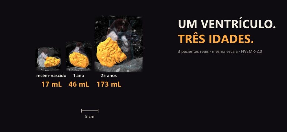
Um recém-nascido sem cirurgia, uma criança de 1 ano depois do Glenn e um adulto de 25 anos depois do Fontan, em ressonâncias reais e na mesma escala. O ventrículo único tem 17, 46 e 173 mL. Ele cresce com o corpo; o que cada cirurgia muda é o trabalho que ele faz.
📅 Novembro de 2026
Navegue pelo Artigo
Três pessoas, uma fisiologia e o que a imagem consegue medir.
- 1. Por que olhar três corações juntos
- 2. Assista: Um ventrículo, três idades
- 3. Passeio 3D: os três corações
- 4. Os três casos, em números
- 5. Recém-nascido: um ventrículo para o corpo e o pulmão
- 6. Um ano: depois do Glenn
- 7. Vinte e cinco anos: depois do Fontan
- 8. O volume cresce; a cirurgia muda o trabalho
- 9. Por que isso importa: os adultos já são maioria
- 10. Como foi feito e o que deu errado
- 11. O que é real e o que é calculado
- 12. Perguntas frequentes
- 13. Considerações Finais
- 14. Referências
1. Por que olhar três corações juntos
Quem cuida de criança com ventrículo único conhece cada etapa de cor: o recém-nascido, o Glenn, o Fontan. Raramente vê as três lado a lado, na mesma régua.
Fui atrás de ressonâncias reais num banco público e encontrei três pessoas com ventrículo único em idades diferentes. Coloquei os corações na mesma escala, em milímetros, e medi o volume de sangue dentro do ventrículo. Não é a mesma pessoa crescendo; são três pacientes, cada um num capítulo da mesma fisiologia, e isso vale para tudo o que vem abaixo.
2. Assista: Um ventrículo, três idades
Cerca de um minuto e vinte, ressonância real e uma voz em português.
3. Passeio 3D: os três corações
Os três corações na mesma escala, lado a lado. Siga os sete passos: cada um acende o que importa, e o teste no fim pede que você toque a estrutura certa no coração certo.
Três corações reais, um ventrículo em cada, em sete passos.
Recém-nascido, 1 ano e 25 anos, lado a lado e na mesma escala. Cada passo acende só o que importa; o que pulsa é o foco. Depois gire, amplie e faça o teste.
O modelo só é baixado quando você abre.
4. Os três casos, em números
Os três vêm do HVSMR-2.0, um banco público de 60 ressonâncias cardíacas com segmentação manual [1]. Os três estão registrados no banco com ventrículo único.
| Caso | Idade | Cirurgia | Ventrículo único | Outras marcas no banco |
|---|---|---|---|---|
| pat58 | recém-nascido | nenhuma | 17 mL | dupla via de saída, átrio comum |
| pat57 | 1 ano | Glenn (bilateral) | 46 mL | duas cavas superiores, átrios invertidos, mesocardia, atresia pulmonar |
| pat56 | 25 anos | Glenn e Fontan | 173 mL | heterotaxia, dextrocardia, duas cavas superiores, átrio comum |
O volume é o sangue dentro da cavidade, contado voxel a voxel na segmentação. Não é o músculo. Do recém-nascido ao adulto, são 10,2 vezes mais.

5. Recém-nascido: um ventrículo para o corpo e o pulmão
Sem cirurgia, um ventrículo só manda sangue para dois circuitos ao mesmo tempo. No pat58, aorta e artéria pulmonar saem da mesma câmara, e o ventrículo tem 17 mL. O fluxo desenhado no filme é esquema sobre a anatomia real.

6. Um ano: depois do Glenn
No Glenn, a cava superior é ligada direto à artéria pulmonar. O sangue da metade de cima do corpo chega ao pulmão sem passar pelo ventrículo. O pat57 tem duas cavas superiores, e as duas abrem na pulmonar: é um Glenn bilateral. O ventrículo tem 46 mL.

7. Vinte e cinco anos: depois do Fontan
Fontan e Baudet descreveram em 1971 uma operação que levava todo o sangue das cavas ao pulmão [2]. No pat56, as duas cavas superiores e a cava inferior chegam à pulmonar; a inferior sobe como um tubo, encostada ao átrio sem se abrir nele, o que é compatível com um conduto extracardíaco. Nenhum sangue das cavas passa pelo ventrículo antes do pulmão. O ventrículo tem 173 mL.
A declaração da American Heart Association sobre a circulação de Fontan descreve o preço desse arranjo: pressão venosa sistêmica cronicamente alta e débito cardíaco reduzido [3].

8. O volume cresce; a cirurgia muda o trabalho
É tentador ler 17, 46 e 173 como efeito das cirurgias. Os dados não deixam: não há peso nem altura no banco, e cada número é de uma pessoa diferente. O volume acompanha o corpo.
O que as cirurgias mudam é o trabalho. No recém-nascido, o ventrículo bombeia para o corpo e para o pulmão. Depois do Glenn, as cavas superiores chegam ao pulmão sem ele. Depois do Fontan, todas as cavas chegam sem ele.
A pergunta do filme
Quando o Fontan falha, o gargalo é o ventrículo ou o circuito sem bomba? Gewillig e colegas resumem a mudança assim: no Fontan, o controle do débito e da congestão sai das mãos do ventrículo e passa a outras forças do circuito [4]. O episódio 3 desta série mostrou esse circuito, sem nenhum milímetro de ventrículo no caminho do sangue até o pulmão.

9. Por que isso importa: os adultos já são maioria
Em Quebec, os adultos eram 66% de todos que viviam com cardiopatia congênita em 2010, e a prevalência de cardiopatia grave entre adultos cresceu 55% de 2000 a 2010 [5]. Corações univentriculares estão dentro dessa definição de grave. A AHA estima até 70 mil pessoas vivas com circulação de Fontan no mundo e uma sobrevida em 30 anos acima de 80% para quem é operado hoje [3].
O adulto de 25 anos do filme faz parte dessa conta, e o cuidado de quem chega a essa idade já não cabe só na cardiologia pediátrica.
10. Como foi feito e o que deu errado
Tirei um quarto caso. O pat50 tem 9 anos e continua em Glenn; numa linha comum de cuidado, o Fontan viria antes disso. Conferi se era um Kawashima (a cava inferior interrompida e drenando pela ázigos), e não é: a cava inferior chega ao átrio comum. Os dados não dizem por que o Fontan não foi feito, e uma escada com esse degrau sem explicação ia confundir mais do que ensinar.
O outro erro foi meu. Para saber quem tinha Glenn, contei os pontos em que a segmentação da cava encosta na da pulmonar. No recém-nascido, essa conta também deu contato. Olhando a junção na própria ressonância, a cava do recém-nascido só encosta a parede na pulmonar, com luzes separadas; na criança de 1 ano, as duas cavas superiores abrem na pulmonar. Contato de rótulos não prova conexão, e o roteiro mudou para dizer “as cavas superiores, aqui são duas”.

11. O que é real e o que é calculado
| Elemento | Origem |
|---|---|
| Cortes da ressonância e superfícies dos corações | Real: HVSMR-2.0, segmentação dos autores do banco |
| Volumes | Calculados: voxels do rótulo ventricular × volume do voxel |
| Escala | Mesma escala em milímetros nos três, sem perspectiva; régua de 5 cm |
| Leitura das conexões das cavas | Leitura de IA sobre a segmentação, conferida nos cortes; revisão por cardiologista pediátrico humano recomendada |
| Fluxo no recém-nascido | Esquema, marcado como tal |
| Voz | Sintética (Gemini TTS); roteiro e revisão do Dr. Mbula |
A morfologia do ventrículo único (esquerdo ou direito) não foi confirmada; o rótulo do banco não prova isso, e o texto não afirma. Uma câmara rudimentar pode existir sem rótulo próprio. Material educativo: as decisões são do médico assistente.
12. Perguntas frequentes
É a mesma criança crescendo?
Não. São três pessoas diferentes, cada uma num momento. O banco é transversal.
Algum deles tem hipoplasia do coração esquerdo?
Nenhum está registrado assim. O banco não tem caso de hipoplasia do coração esquerdo.
Os volumes servem como referência?
Não. Sem peso e altura, não há indexação, e são três casos.
A IA fez o diagnóstico?
Não. Os diagnósticos vêm do banco e a segmentação é dos autores. Os scripts medem o que a segmentação mostra; a leitura das conexões é de IA, foi conferida nos cortes e tem os erros expostos acima.
Quer ensinar anatomia assim?
A Metodologia AIMED organiza o uso de IA em medicina, com revisão clínica em cada etapa.
13. Considerações Finais
Três corações reais, um ventrículo em cada um, 17, 46 e 173 mL. O número cresce com o corpo, e a cirurgia tira do ventrículo uma parte do trabalho de cada vez.
💡 Connecting the Dots. O adulto de 25 anos é a parte que mais me interessa. Chegou lá com um ventrículo só, e é por pessoas assim que os adultos já são maioria entre quem vive com cardiopatia congênita. Quando esse Fontan começar a falhar, a pergunta do filme deixa de ser exercício: o problema estará nos 173 mL ou no circuito que trabalha sem bomba?
Leia também Cava superior esquerda em 3D, o episódio anterior, e Bandagem da pulmonar em 3D, que mostra uma das primeiras etapas do mesmo caminho.
Este episódio só existe porque três pessoas, do primeiro dia de vida aos 25 anos, estão numa base pública.
Referências
- Pace DF, Contreras HTM, Romanowicz J, et al. HVSMR-2.0: A 3D cardiovascular MR dataset for whole-heart segmentation in congenital heart disease. Sci Data. 2024;11:721. PMID: 38956063
- Fontan F, Baudet E. Surgical repair of tricuspid atresia. Thorax. 1971;26(3):240-8. PMID: 5089489. PMCID: PMC1019078
- Rychik J, Atz AM, Celermajer DS, et al. Evaluation and Management of the Child and Adult With Fontan Circulation: A Scientific Statement From the American Heart Association. Circulation. 2019;140(6):e234-e284. PMID: 31256636
- Gewillig M, Brown SC, van de Bruaene A, Rychik J. Providing a framework of principles for conceptualising the Fontan circulation. Acta Paediatr. 2020;109(4):651-658. PMID: 31737940. PMCID: PMC7155129
- Marelli AJ, Ionescu-Ittu R, Mackie AS, Guo L, Dendukuri N, Kaouache M. Lifetime prevalence of congenital heart disease in the general population from 2000 to 2010. Circulation. 2014;130(9):749-56. PMID: 24944314
Dados: HVSMR-2.0, CC BY 4.0, modificado (superfícies, cortes e medidas). Música do filme: ‘Amberlight’ by Scott Buckley – released under CC-BY 4.0. www.scottbuckley.com.au. Voz sintética (Gemini TTS: Zubenelgenubi).
One ventricle, three ages: the single ventricle in real hearts
A newborn with no surgery, a 1-year-old after a Glenn and a 25-year-old after a Fontan, on real MRI scans and on the same scale. The single ventricle holds 17, 46 and 173 mL. It grows with the body; what each operation changes is the work it does.
📅 November 2026
Navigate the Article
Three people, one physiology and what the images can measure.
- 1. Why look at three hearts together
- 2. Watch: One ventricle, three ages
- 3. 3D tour: the three hearts
- 4. The three cases, in numbers
- 5. Newborn: one ventricle for the body and the lungs
- 6. One year: after the Glenn
- 7. Twenty-five years: after the Fontan
- 8. The volume grows; surgery changes the workload
- 9. Why it matters: adults are now the majority
- 10. How it was made and what went wrong
- 11. What is real and what is calculated
- 12. Frequently asked questions
- 13. Final Thoughts
- 14. References
1. Why look at three hearts together
Anyone who cares for children with a single ventricle knows each stage by heart: the newborn, the Glenn, the Fontan. They rarely see all three side by side, on the same ruler.
I looked for real MRI scans in a public dataset and found three people with a single ventricle at different ages. I put the hearts on the same millimetre scale and measured the blood volume inside the ventricle. This is not one person growing up; they are three patients, each in one chapter of the same physiology, and that holds for everything below.
2. Watch: One ventricle, three ages
About a minute and twenty seconds, real MRI and one voice in English.
3. 3D tour: the three hearts
The three hearts at the same scale, side by side. Follow the seven steps: each one lights what matters, and the test at the end asks you to tap the right structure in the right heart.
Three real hearts, one ventricle each, in seven steps.
Newborn, age 1 and age 25, side by side at the same scale. Each step lights only what matters; what pulses is the focus. Then rotate, zoom and take the test.
The model downloads only when you open it.
4. The three cases, in numbers
All three come from HVSMR-2.0, a public dataset of 60 cardiac MRI scans with manual segmentation [1]. All three are recorded in the dataset with a single ventricle.
| Case | Age | Surgery | Single ventricle | Other dataset labels |
|---|---|---|---|---|
| pat58 | newborn | none | 17 mL | double outlet, common atrium |
| pat57 | 1 year | Glenn (bilateral) | 46 mL | two superior cavae, inverted atria, mesocardia, pulmonary atresia |
| pat56 | 25 years | Glenn and Fontan | 173 mL | heterotaxy, dextrocardia, two superior cavae, common atrium |
The volume is the blood inside the cavity, counted voxel by voxel in the segmentation. It is not the muscle. From the newborn to the adult, it is 10.2 times larger.

5. Newborn: one ventricle for the body and the lungs
Without surgery, one ventricle pumps into two circuits at once. In pat58, the aorta and the pulmonary artery leave the same chamber, and the ventricle holds 17 mL. The flow drawn in the film is a schematic over the real anatomy.

6. One year: after the Glenn
In a Glenn, the superior vena cava is connected straight to the pulmonary artery. Blood from the upper half of the body reaches the lungs without passing the ventricle. Pat57 has two superior cavae, and both open into the pulmonary artery: a bilateral Glenn. The ventricle holds 46 mL.

7. Twenty-five years: after the Fontan
In 1971, Fontan and Baudet described an operation that sent all the caval blood to the lungs [2]. In pat56, both superior cavae and the inferior cava reach the pulmonary artery; the inferior one runs up as a tube, touching the atrium without opening into it, which fits an extracardiac conduit. No caval blood passes through the ventricle before the lungs. The ventricle holds 173 mL.
The American Heart Association statement on the Fontan circulation describes the price of this arrangement: chronically high systemic venous pressure and reduced cardiac output [3].

8. The volume grows; surgery changes the workload
It is tempting to read 17, 46 and 173 as the effect of surgery. The data don’t allow it: there is no weight or height in the dataset, and each number belongs to a different person. The volume follows the body.
What surgery changes is the workload. In the newborn, the ventricle pumps to the body and the lungs. After the Glenn, the superior cavae reach the lungs without it. After the Fontan, every cava does.
The film’s question
When a Fontan fails, is the bottleneck the ventricle or the circuit without a pump? Gewillig and colleagues put the shift this way: in a Fontan, control of cardiac output and congestion is taken away from the ventricle and handed to other forces in the circuit [4]. Episode 3 of this series showed that circuit, with not one millimetre of ventricle on the blood’s path to the lungs.

9. Why it matters: adults are now the majority
In Quebec, adults were 66% of everyone living with congenital heart disease in 2010, and the prevalence of severe disease among adults rose 55% from 2000 to 2010 [5]. Univentricular hearts fall within that definition of severe. The AHA estimates up to 70,000 people alive with a Fontan circulation worldwide and a 30-year survival above 80% for those operated on today [3].
The 25-year-old in the film is part of that count, and care at that age no longer fits in paediatric cardiology alone.
10. How it was made and what went wrong
I left out a fourth case. Pat50 is 9 years old and still in Glenn; on a usual pathway, the Fontan would have come earlier. I checked whether it was a Kawashima (an interrupted inferior cava draining through the azygos), and it is not: the inferior cava reaches the common atrium. The data don’t say why the Fontan wasn’t done, and a ladder with that unexplained step would confuse more than teach.
The other mistake was mine. To find out who had a Glenn, I counted the points where the caval segmentation touches the pulmonary one. In the newborn, that count also showed contact. Looking at the junction on the MRI itself, the newborn’s cava only touches the pulmonary artery wall, with separate lumens; in the 1-year-old, both superior cavae open into the pulmonary artery. Touching labels don’t prove a connection, and the script changed to say “the superior cavae, two of them here”.

11. What is real and what is calculated
| Element | Source |
|---|---|
| MRI slices and heart surfaces | Real: HVSMR-2.0, segmentation by the dataset authors |
| Volumes | Calculated: voxels of the ventricular label × voxel volume |
| Scale | Same millimetre scale for all three, no perspective; 5 cm bar |
| Reading of the caval connections | AI reading of the segmentation, checked on the slices; review by a human paediatric cardiologist recommended |
| Flow in the newborn | Schematic, labelled as such |
| Voice | Synthetic (Gemini TTS); script and review by Dr. Mbula |
The morphology of the single ventricle (left or right) was not confirmed; the dataset label does not prove it, and the text does not claim it. A rudimentary chamber may exist without its own label. Educational material: decisions belong to the treating physician.
12. Frequently asked questions
Is it the same child growing up?
No. They are three different people, each at one moment. The dataset is cross-sectional.
Does any of them have hypoplastic left heart syndrome?
None is recorded that way. The dataset has no case of hypoplastic left heart.
Can the volumes be used as reference values?
No. Without weight and height there is no indexing, and there are only three cases.
Did AI make the diagnosis?
No. The diagnoses come from the dataset and the segmentation from its authors. The scripts measure what the segmentation shows; the reading of the connections is by AI, was checked on the slices and has its mistakes shown above.
Want to teach anatomy like this?
The AIMED Methodology organises the use of AI in medicine, with clinical review at every step.
13. Final Thoughts
Three real hearts, one ventricle in each, 17, 46 and 173 mL. The number grows with the body, and surgery takes part of the work away from the ventricle, one step at a time.
💡 Connecting the Dots. The 25-year-old is the part that interests me most. This patient got there with a single ventricle, and people like this are why adults are now the majority of those living with congenital heart disease. When this Fontan begins to fail, the film’s question stops being an exercise: will the problem be in the 173 mL or in the circuit working without a pump?
Read also Left superior vena cava in 3D, the previous episode, and Pulmonary artery banding in 3D, one of the first steps on the same path.
This episode exists only because three people, from their first day of life to age 25, are in a public dataset.
References
- Pace DF, Contreras HTM, Romanowicz J, et al. HVSMR-2.0: A 3D cardiovascular MR dataset for whole-heart segmentation in congenital heart disease. Sci Data. 2024;11:721. PMID: 38956063
- Fontan F, Baudet E. Surgical repair of tricuspid atresia. Thorax. 1971;26(3):240-8. PMID: 5089489. PMCID: PMC1019078
- Rychik J, Atz AM, Celermajer DS, et al. Evaluation and Management of the Child and Adult With Fontan Circulation: A Scientific Statement From the American Heart Association. Circulation. 2019;140(6):e234-e284. PMID: 31256636
- Gewillig M, Brown SC, van de Bruaene A, Rychik J. Providing a framework of principles for conceptualising the Fontan circulation. Acta Paediatr. 2020;109(4):651-658. PMID: 31737940. PMCID: PMC7155129
- Marelli AJ, Ionescu-Ittu R, Mackie AS, Guo L, Dendukuri N, Kaouache M. Lifetime prevalence of congenital heart disease in the general population from 2000 to 2010. Circulation. 2014;130(9):749-56. PMID: 24944314
Data: HVSMR-2.0, CC BY 4.0, modified (surfaces, slices and measurements). Film music: ‘Amberlight’ by Scott Buckley – released under CC-BY 4.0. www.scottbuckley.com.au. Synthetic voice (Gemini TTS: Zubenelgenubi).
◆ Novidades
- Cateter à esquerda da coluna: mal posicionado ou cava esquerda?NOVO
- Por que apertar a artéria de um bebê? A bandagem da pulmonar num coração real
- Heterotaxia em 3D: a veia cava inferior entrou do outro lado, e isso não diz qual átrio é qual