
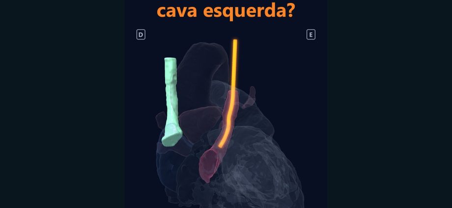
Cateter à esquerda da coluna: mal posicionado ou cava esquerda?
Na ressonância real de um adulto de 27 anos com duas cavas superiores, a esquerda corre a 4,9 cm da direita e chega ao átrio direito pelo seio coronário, que mede 13 mm por dentro, mais largo que cada uma das cavas. A imagem mostra o caminho. Se o teto do seio está inteiro, ela não consegue dizer, e é isso que decide se um cateter ali pode levar ar ou coágulo para a circulação sistêmica.
📅 Novembro de 2026
Navegue pelo Artigo
Do telefonema do leito às medidas, com o que a imagem não alcança.
- 1. Por que isso importa no plantão
- 2. Assista: Mal posicionado ou cava esquerda?
- 3. Passeio 3D: as duas cavas
- 4. O que é a cava superior esquerda persistente
- 5. O coração real, em números
- 6. O seio coronário medido
- 7. O que conferir antes de usar o cateter
- 8. Se não for cava esquerda: o diferencial
- 9. Para onde ela drena
- 10. Se for usar, onde fica a ponta
- 11. Traduzindo para o chão da UTI
- 12. Como foi feito e o que deu errado
- 13. O que é real e o que é calculado
- 14. Perguntas frequentes
- 15. Considerações Finais
- 16. Referências
1. Por que isso importa no plantão
Três da manhã. A residente liga do leito: o cateter entrou pelo lado esquerdo, e a radiografia mostra o fio descendo à esquerda da coluna, reto, colado à borda do coração. A pergunta chega pronta. Tira e passa de novo, ou é uma cava esquerda?
A dúvida tem fundamento. Na cava superior esquerda persistente, o cateter desce justamente pela borda esquerda do mediastino e pode ser confundido com mau posicionamento [1]. O contrário também acontece, e um fio nesse lugar pode estar numa artéria, numa veia pequena do tórax ou fora do vaso.
Fui atrás de uma ressonância real com duas cavas superiores, num banco público, e medi o que ela permite medir. Onde a imagem parou de responder, deixei escrito.
2. Assista: Mal posicionado ou cava esquerda?
Cerca de um minuto e meio, ressonância real e duas vozes em português: a residente que liga do leito e o plantonista que responde. A primeira medida aparece antes dos 15 segundos.
3. Passeio 3D: as duas cavas
Os dois corações na mesma escala: o adulto com duas cavas superiores e o de referência, com 4 anos, que tem uma só. Gire o modelo e siga a cava esquerda por trás do coração até o seio coronário.
Siga a cava esquerda até o seio coronário, em sete passos.
Dois corações reais, lado a lado e na mesma escala. Cada passo acende só o que importa; a cava esquerda pulsa em vinho. Depois gire, amplie e faça o teste.
O modelo só é baixado quando você abre.
4. O que é a cava superior esquerda persistente
É uma segunda veia cava superior, do lado esquerdo. Na maioria das vezes ela desce atrás do coração e chega ao átrio direito pelo seio coronário [2]. Quase sempre a cava direita também existe, e então são duas, como no caso deste episódio.
A frequência depende de quem se olha. A revisão de Povoski e Khabiri cita 0,3 a 0,5% na população geral e até 12% em cardiopatia congênita [2]. Roldan e Paniagua dão 0,3% em pessoas saudáveis e 4,3% em cardiopatia [3]. Numa série de 4.000 crianças operadas do coração, a cava esquerda apareceu em 6,5% [4]. O filme não cita prevalência, porque as séries variam demais para caber numa legenda.
Tradutor de siglas, pela pergunta do plantão
“Onde esse fio foi parar?” VCS: veia cava superior. Cava esquerda (em inglês, left SVC ou PLSVC): a segunda cava, do lado esquerdo. Seio coronário: a veia que corre no sulco atrás do coração, entre o átrio e o ventrículo esquerdos, e desemboca no átrio direito. AD e AE: átrio direito e átrio esquerdo. PICC: cateter central de inserção periférica, passado pelo braço.

5. O coração real, em números
O caso é o pat7 do banco público HVSMR-2.0: um adulto de 27 anos, registrado com cava superior bilateral e sem outra cardiopatia marcada no banco. A comparação é o pat5, uma criança de 4 anos com coração normal e uma só cava superior, à direita. A anatomia é real e anonimizada; a história do cateter é ilustrativa.
| Medida | Adulto, 27 anos (pat7) | Referência, 4 anos (pat5) | Leitura |
|---|---|---|---|
| Cavas superiores | duas, direita e esquerda | uma, à direita | → cava bilateral |
| Distância lateral entre os centros das cavas | 4,9 cm (mediana de 67 cortes; 4,4–5,3 cm) | não se aplica | → duas cavas separadas |
| Diâmetro inscrito em 3D, cava esquerda (trecho superior) | 7–8 mm | não existe | → calibre de cava |
| Diâmetro inscrito em 3D, cava direita | 8,5–9,5 mm | não comparado | → calibre de cava |
| Diâmetro inscrito em 3D, seio coronário | 12,4–13,1 mm | não comparado | ↑ mais largo que cada cava |
| Altura do trecho segmentado da cava esquerda | 81 mm | não existe | → do topo do recorte ao seio |
| Contato da cava esquerda com AD · AE · tronco pulmonar · aorta (voxels) | 1.454 · 50 · 95 · 0 | não se aplica | → chega ao AD |
| Menor distância do seio ao AE · ao AD | 1,0 mm · 0,7 mm | não se aplica | ⚠ teto não avaliável |
Voxel de 0,75 × 0,73 × 0,73 mm. O contato foi contado com a máscara dilatada em 2 voxels: mostra vizinhança anatômica entre as máscaras e não mede fluxo nem o tamanho de uma passagem. Os 50 voxels junto ao AE são a mesma proximidade de 1,0 mm que aparece na última linha.

6. O seio coronário medido
A cava esquerda desce ao lado do tronco pulmonar e da aurícula esquerda, entra no sulco atrioventricular posterior, entre o átrio e o ventrículo esquerdos, e chega ao átrio direito pela parte posterior e inferior. É o trajeto típico de um seio coronário que recebe uma cava esquerda.
Para medir o calibre, uma esfera cresce dentro da máscara do vaso até tocar as paredes. O diâmetro dessa maior esfera é o diâmetro inscrito em 3D, que não depende da direção em que o vaso foi cortado. Com ele, a cava esquerda tem cerca de 8 mm no trecho superior, a direita cerca de 9 mm e o seio coronário 13 mm. O seio é mais largo que cada uma das cavas.
Diâmetro inscrito, pela pergunta do plantão
“Quanto esse vaso tem por dentro?” O diâmetro inscrito é o tamanho da maior bola que cabe no lúmen. Um vaso deitado, cortado de lado, não engana esse número.
Um exemplo concreto: “o seio de mais de um centímetro”
No relato de Dontukurthy e colegas, um seio coronário acima de 1 cm é o sinal que sugere cava esquerda [5]. O seio deste adulto tem 13 mm na ressonância. No eco de beira de leito o número viria de outra janela e de outra borda, e o limiar continua sendo de relato de caso.

7. O que conferir antes de usar o cateter
O plantonista do filme começa pelo exame que pode encerrar a conversa: o sangue. Gasometria venosa e curva de pressão venosa afastam a punção arterial; se for artéria, a dúvida acaba ali. Na série de oito neonatos com PICC na cava esquerda, a gasometria venosa fez parte da confirmação [6]. Num relato, foi a própria curva de pressão, com aspecto “ventricularizado” enquanto a porta do cateter estava dentro do seio coronário, que levantou a suspeita de cava esquerda [5].
Depois, o trajeto. A cava esquerda desce reta pela borda esquerda do mediastino e só lá embaixo vira para dentro, rumo ao átrio direito [1]. A radiografia sozinha muitas vezes não basta para definir a posição [7], e contraste ou tomografia ajudam a confirmar [3].
No eco, a pista é o seio coronário dilatado [8]. Microbolhas injetadas numa veia do braço esquerdo opacificam o seio antes do átrio direito [8, 5].
⚠ Ressalva
Nenhum desses exames existe para este adulto: a base pública traz só a ressonância. O eco do filme é esquema, com o selo “não é deste paciente”. E a vista frontal do filme é projeção da segmentação da ressonância, não radiografia.

8. Se não for cava esquerda: o diferencial
Um cateter à esquerda da coluna também pode estar onde não deveria. Os relatos e revisões citam artéria (carótida, aorta) e posição extraluminal [10]; veia torácica interna, hemiázigo acessória e pericardiofrênica, sobretudo com punção pela jugular esquerda [7]; e veia intercostal superior esquerda [9].
Várias dessas veias correm do mesmo lado, perto da borda do coração. Por isso o trajeto precisa ser seguido até o fim: a cava esquerda desce até o sulco atrás do coração e vira para o átrio direito [1].
9. Para onde ela drena
Esta é a pergunta que muda a conduta. Na revisão de Povoski e Khabiri, 80 a 92% das cavas esquerdas drenam no átrio direito pelo seio coronário, e 10 a 20% no átrio esquerdo [2]. Na série de Toprak e colegas, com crianças operadas de cardiopatia congênita, 92,3% drenavam para o seio coronário e 7,7% para o átrio esquerdo [4].
Quando a cava esquerda chega ao átrio esquerdo, por exemplo por um seio coronário sem teto, o sangue dela entra direto na circulação sistêmica. Ar ou coágulo vindos do cateter seguem o mesmo caminho, e o nome disso é embolia paradoxal [2]. A mesma revisão recomenda confirmar a drenagem antes de usar o acesso [2].
Neste caso
Na segmentação, a cava esquerda chega ao átrio direito pelo seio coronário, com 1.454 voxels de contato com o AD. O teto do seio não é avaliável: a menor distância entre a máscara do seio e a do átrio esquerdo é de 1,0 mm, perto de um voxel e meio. Nesta resolução, não dá para afirmar nem excluir seio sem teto.

10. Se for usar, onde fica a ponta
Não encontrei diretriz formal sobre a posição da ponta na cava esquerda. Há séries e relatos. Na série de Chen e colegas, oito neonatos receberam PICC com a ponta no terço médio ou inferior da cava esquerda, sem complicações [6], e a revisão de Povoski e Khabiri apoia conduta semelhante [2].
Dentro do seio coronário, a lista de problemas relatados é longa: arritmia, choque, tamponamento e trombose do seio [8], além de perfuração e medidas erradas [11]. Há também relato de cateter que entrou no seio coronário pela cava esquerda [10].
⚠ Sem diretriz formal
Oito neonatos e relatos de caso não sustentam recomendação forte. O filme mostra “sem diretriz formal” na tela pelo mesmo motivo.
11. Traduzindo para o chão da UTI
A ressonância deste episódio mostra o caminho de uma cava esquerda real. À beira do leito, a confirmação é feita no paciente.
- ☐ Sangue venoso confirmado por gasometria e curva de pressão.
- ☐ Trajeto que desce reto pela borda esquerda do mediastino e só embaixo vira para dentro [1]; na dúvida, contraste ou tomografia [3].
- ☐ Eco com seio coronário largo e microbolhas pelo braço esquerdo [8, 5].
- ☐ Drenagem conhecida antes de usar o acesso, porque a minoria que vai para o átrio esquerdo carrega o risco de embolia paradoxal [2].
- ☐ Ponta fora do seio coronário [6, 8].
Material educativo. As decisões são sempre do médico assistente e da equipe que conhece o paciente.
12. Como foi feito e o que deu errado
A triagem nos cortes foi feita em 12 de novembro de 2026, sobre a segmentação dos autores do banco. Errei antes de chegar a estes números, e conto os erros porque eles explicam as medidas:
- Dezoito milímetros que eram treze. Minha primeira conta do seio coronário deu 18,2 mm. Eu tinha convertido em diâmetro a área do vaso no corte axial. Só que o seio corre quase deitado no sulco atrioventricular, e o corte axial o pega de lado, como uma fatia oblíqua de salsicha. Medido por dentro, com a maior esfera que cabe em 3D, deu 13 mm.
- Isolada, não: bilateral. Na minha nota de retomada, o caso estava como “cava esquerda isolada”. A direita existe. “Isolada” só vale no sentido de não haver outra cardiopatia marcada no banco.
- A régua. Para a tela dizer 4,9 cm sem contradizer a tabela, a régua entre as cavas foi posta num corte em que a distância é igual à mediana dos 67 cortes, 48,6 mm.
- Um estreitamento que ficou de fora. Num trecho de cerca de 10 mm, a cava esquerda afina para 3,3 a 4,4 mm na segmentação. Pode ser compressão, pode ser a borda da máscara. Não sei, e por isso o número não entrou no filme.
- A frase das cavas. Na narração em português, o plantonista diz que o seio é “mais largo que as duas cavas”. Quer dizer mais largo que cada uma delas (13 mm contra 8 e 9 mm), e não que a soma.

13. O que é real e o que é calculado
| Elemento | Origem |
|---|---|
| Cortes da ressonância e superfícies dos corações | Real: HVSMR-2.0, segmentação dos autores do banco, re-rotulada aqui para separar a cava esquerda (com o seio) da direita |
| Distâncias e diâmetros | Calculados sobre a segmentação: distância lateral entre centros nos cortes axiais; diâmetro inscrito em 3D |
| Leitura do trajeto e da drenagem | Leitura de IA sobre a segmentação, no papel de ecocardiografista; revisão por cardiologista pediátrico humano recomendada |
| Vista frontal do filme | Projeção da segmentação, não radiografia |
| Fio do cateter, curva de pressão, eco e microbolhas | Esquemas, marcados como tal |
| História clínica | Ilustrativa |
| Vozes | Sintéticas (Gemini TTS: Kore e Zubenelgenubi); roteiro e revisão do Dr. Mbula |
O banco não tem rótulo próprio para o seio coronário: ele veio junto com a cava, e o segmento largo do sulco foi lido como seio. A ponte venosa entre as cavas (a veia inominada) não aparece porque o recorte termina antes; isso não quer dizer que ela falte. As referências 1, 4 e 11 foram lidas pelo resumo, e as demais no texto integral. Caso clínico ilustrativo e material educativo: as decisões são do médico assistente.
14. Perguntas frequentes
Este adulto tem seio coronário sem teto?
Não sei. A separação de 1,0 mm do átrio esquerdo fica perto de um voxel e meio, e nesta resolução não dá para afirmar nem excluir.
Os 13 mm valem para o eco?
Não diretamente. São o diâmetro inscrito na máscara da ressonância. O eco mede por outra janela, com outra borda, e o limiar de 1 cm vem de um relato de caso [5].
Posso deixar a ponta na cava esquerda?
Há série neonatal com a ponta no terço médio ou inferior da cava esquerda [6], sem diretriz formal. A decisão é da equipe, depois de confirmar sangue venoso e drenagem [2].
A IA fez o diagnóstico?
Não. O registro de cava bilateral vem do banco, e a segmentação é dos autores. Os scripts medem o que a segmentação mostra; a leitura do trajeto é de IA, foi revisada e tem os erros expostos acima. A revisão por cardiologista pediátrico humano continua recomendada.
Quer ensinar anatomia assim?
A Metodologia AIMED organiza o uso de IA em medicina, com revisão clínica em cada etapa.
15. Considerações Finais
“Mal posicionado ou cava esquerda?” Neste exame, a resposta é cava esquerda: ela existe, desce pela borda do coração e chega ao átrio direito por um seio de 13 mm. Com um cateter dentro, porém, a pergunta seguinte é outra, e a imagem não chega até ela.
💡 Connecting the Dots. Quase todo texto sobre cava esquerda repete que ela drena no átrio direito na maioria dos casos. É verdade, e as séries dão 80 a 92% [2]. O número que pesa à beira do leito é o resto: a minoria que chega ao átrio esquerdo, por onde ar ou coágulo do cateter vão direto para a circulação sistêmica. Numa ressonância boa, o teto do seio deste adulto ficou a 1,0 mm do átrio esquerdo, um voxel e meio, e eu não consegui fechá-lo. Medi o trajeto e o calibre e deixei escrito onde a imagem parou. Se o cateter pode ser usado, quem responde é a confirmação feita no paciente, com gasometria, curva de pressão, trajeto e eco com microbolhas pelo braço esquerdo.
Leia também os episódios Bandagem da pulmonar em 3D, o anterior, e Heterotaxia em 3D, sobre a outra cava, a inferior.
Este exame só pôde ser estudado porque a ressonância de um adulto de 27 anos está numa base pública.
Referências
- Schummer W, Schummer C, Hoffmann E, Gerold M. Persistent left superior vena cava: clinical implications for central venous cannulation. Nutr Clin Pract. 2002;17(5):304-8. PMID: 16215005
- Povoski SP, Khabiri H. Persistent left superior vena cava: review of the literature, clinical implications, and relevance of alterations in thoracic central venous anatomy as pertaining to the general principles of central venous access device placement and venography in cancer patients. World J Surg Oncol. 2011;9:173. PMID: 22204758. PMCID: PMC3266648
- Roldan CJ, Paniagua L. Central Venous Catheter Intravascular Malpositioning: Causes, Prevention, Diagnosis, and Correction. West J Emerg Med. 2015;16(5):658-64. PMID: 26587087. PMCID: PMC4644031
- Toprak MHH, Serifoglu S, Tanıdır İC, et al. Frequency of Persistent Left Superior Vena Cava and Its Impact on Outcomes in Children Undergoing Congenital Heart Surgery. Braz J Cardiovasc Surg. 2025;40(6):e20240446. PMID: 41217972
- Dontukurthy S, Yamaguchi Y, Tobias JD. Persistent Left Superior Vena Cava Suggested by an Unusual Central Venous Pressure Waveform. Thorac Cardiovasc Surg Rep. 2020;9(1):e15-e17. PMID: 32355604. PMCID: PMC7188515
- Chen Q, Hu YL, Li YX, Huang X. Peripherally inserted central catheter placement in neonates with persistent left superior vena cava: Report of eight cases. World J Clin Cases. 2021;9(26):7944-7953. PMID: 34621850. PMCID: PMC8462248
- Ghatak T, Gupta B, Ghatak D, Samanta S, Nachimuthu N. Anatomical patterns and clinical outcomes of central venous catheter malpositions: a systematic review of published case reports and case series. Indian J Anaesth. 2026;70(Suppl 3):S180-S193. PMID: 42819636. PMCID: PMC13626507
- Goyal SK, Punnam SR, Verma G, Ruberg FL. Persistent left superior vena cava: a case report and review of literature. Cardiovasc Ultrasound. 2008;6:50. PMID: 18847480. PMCID: PMC2576163
- Padovan RS, Paar MH, Aurer I. (Mis)placed central venous catheter in the left superior intercostal vein. Radiol Oncol. 2011;45(1):27-30. PMID: 22933931. PMCID: PMC3423719
- Syska B, Veer AS, Matusik PS, et al. Malposition of Central Venous Catheter into Coronary Sinus throughout the Persistent Left Superior Vena Cava… Diagnostics (Basel). 2024;14(10):1038. PMID: 38786336. PMCID: PMC11119990
- Atoa SM, Berry-Rieser AD, Vitin AA, et al. Inadvertent Coronary Sinus Cannulation During Central Venous Catheter Placement in a Liver Transplant Recipient with PLSVC. Transplant Proc. 2026 (ahead of print). PMID: 42864219
Dados: HVSMR-2.0 (Pace DF, Contreras HTM, Romanowicz J, et al. Sci Data. 2024;11:721. PMID: 38956063), CC BY 4.0, modificado (segmentação re-rotulada, com a cava esquerda e o seio separados da cava direita; superfícies; medidas). Música do filme: ‘Memories Of Stone’ by Scott Buckley – released under CC-BY 4.0. www.scottbuckley.com.au. Vozes sintéticas (Gemini TTS: Kore e Zubenelgenubi).
Line left of the spine: misplaced or a left SVC?
In the real MRI of a 27-year-old with two superior venae cavae, the left one runs 4.9 cm from the right and reaches the right atrium through the coronary sinus, which is 13 mm across on the inside, wider than either cava. The image shows the path. Whether the roof of the sinus is intact, it cannot say, and that is what decides whether a line placed there could carry air or clot into the systemic circulation.
📅 November 2026
Navigate the Article
From the call from the bedside to the measurements, and what the image cannot reach.
- 1. Why this matters on the night shift
- 2. Watch: Misplaced or a left SVC?
- 3. 3D tour: the two cavae
- 4. What a persistent left superior vena cava is
- 5. The real heart, in numbers
- 6. The coronary sinus, measured
- 7. What to check before using the line
- 8. If it is not a left SVC: the differential
- 9. Where it drains
- 10. If you use it, where the tip goes
- 11. Translating to the ICU floor
- 12. How it was made and what went wrong
- 13. What is real and what is calculated
- 14. Frequently asked questions
- 15. Final Considerations
- 16. References
1. Why this matters on the night shift
Three in the morning. The resident calls from the bedside: the line went in on the left, and the X-ray shows it running down left of the spine, straight, hugging the heart border. The question comes ready-made. Pull it and try again, or is it a left SVC?
The doubt is well founded. With a persistent left superior vena cava, the catheter runs down exactly along the left mediastinal border and can be mistaken for a malposition [1]. The reverse also happens, and a line in that spot may be in an artery, in a small thoracic vein or outside the vessel.
I went looking for a real MRI with two superior venae cavae, in a public dataset, and measured what it allows. Where the image stopped answering, I wrote it down.
2. Watch: Misplaced or a left SVC?
About a minute and a half, real MRI and two voices in English: the resident calling from the bedside and the attending who answers. The first measurement appears before the 15-second mark.
3. 3D tour: the two cavae
Both hearts at the same scale: the adult with two superior venae cavae and the reference heart, age 4, which has only one. Turn the model and follow the left cava behind the heart into the coronary sinus.
Follow the left cava into the coronary sinus, in seven steps.
Two real hearts, side by side at the same scale. Each step lights only what matters; the left cava pulses in wine. Then rotate, zoom and take the test.
The model downloads only when you open it.
4. What a persistent left superior vena cava is
It is a second superior vena cava, on the left. Most of the time it runs down behind the heart and reaches the right atrium through the coronary sinus [2]. The right cava is usually there as well, so there are two, as in this episode’s case.
How common it is depends on who you look at. The review by Povoski and Khabiri gives 0.3 to 0.5% in the general population and up to 12% in congenital heart disease [2]. Roldan and Paniagua give 0.3% in healthy people and 4.3% in heart disease [3]. In a series of 4,000 children undergoing heart surgery, a left SVC was found in 6.5% [4]. The film quotes no prevalence, because the series vary too much to fit in a caption.
Acronym translator, by the night-shift question
“Where did that line end up?” SVC: superior vena cava. Left SVC (or PLSVC): the second cava, on the left. Coronary sinus: the vein that runs in the groove behind the heart, between the left atrium and left ventricle, and empties into the right atrium. RA and LA: right atrium and left atrium. PICC: peripherally inserted central catheter, placed through the arm.

5. The real heart, in numbers
The case is pat7 from the public HVSMR-2.0 dataset: a 27-year-old adult, recorded with a bilateral superior vena cava and no other heart defect flagged in the dataset. The comparison is pat5, a 4-year-old with a normal heart and a single superior vena cava, on the right. The anatomy is real and anonymised; the line story is illustrative.
| Measurement | Adult, age 27 (pat7) | Reference, age 4 (pat5) | Reading |
|---|---|---|---|
| Superior venae cavae | two, right and left | one, on the right | → bilateral SVC |
| Lateral distance between the centres of the cavae | 4.9 cm (median of 67 slices; 4.4–5.3 cm) | not applicable | → two separate cavae |
| 3D inscribed diameter, left cava (upper segment) | 7–8 mm | absent | → caval calibre |
| 3D inscribed diameter, right cava | 8.5–9.5 mm | not compared | → caval calibre |
| 3D inscribed diameter, coronary sinus | 12.4–13.1 mm | not compared | ↑ wider than either cava |
| Height of the segmented left cava | 81 mm | absent | → from the top of the crop to the sinus |
| Left cava contact with RA · LA · pulmonary trunk · aorta (voxels) | 1,454 · 50 · 95 · 0 | not applicable | → reaches the RA |
| Shortest distance from the sinus to the LA · to the RA | 1.0 mm · 0.7 mm | not applicable | ⚠ roof not assessable |
Voxel size 0.75 × 0.73 × 0.73 mm. Contact was counted with the mask dilated by 2 voxels: it shows anatomical proximity between masks and does not measure flow or the size of a passage. The 50 voxels next to the LA are the same 1.0 mm proximity shown in the last row.

6. The coronary sinus, measured
The left cava runs down beside the pulmonary trunk and the left atrial appendage, enters the posterior atrioventricular groove between the left atrium and left ventricle, and reaches the right atrium from behind and below. That is the typical course of a coronary sinus receiving a left SVC.
To measure calibre, a sphere grows inside the vessel mask until it touches the walls. The diameter of that largest sphere is the 3D inscribed diameter, and it does not depend on the direction in which the vessel was cut. By this measure, the left cava is about 8 mm in its upper segment, the right one about 9 mm and the coronary sinus 13 mm. The sinus is wider than either cava.
Inscribed diameter, by the night-shift question
“How wide is this vessel on the inside?” The inscribed diameter is the size of the largest ball that fits in the lumen. A vessel lying flat and cut at an angle cannot fool this number.
A concrete example: “the sinus over a centimetre”
In the report by Dontukurthy and colleagues, a coronary sinus over 1 cm is the sign that suggests a left SVC [5]. This adult’s sinus is 13 mm on MRI. A bedside echo would get its number from another window and another edge, and the threshold still comes from a case report.

7. What to check before using the line
The attending in the film starts with the test that can end the conversation: the blood. A venous gas and a venous pressure trace rule out arterial puncture; if it is arterial, that settles it. In the series of eight neonates with a PICC in the left SVC, a venous gas was part of the confirmation [6]. In one report, it was the pressure trace itself, looking “ventricularised” while the catheter port sat inside the coronary sinus, that raised the suspicion of a left SVC [5].
Then, the course. A left SVC runs straight down the left mediastinal border and only turns inward at the bottom, toward the right atrium [1]. The X-ray alone is often not enough to define the position [7], and contrast or CT help confirm it [3].
On echo, the clue is a dilated coronary sinus [8]. Bubbles injected into a left arm vein opacify the sinus before the right atrium [8, 5].
⚠ Caveat
None of these tests exist for this adult: the public dataset only holds the MRI. The echo in the film is a schematic, labelled “not this patient”. And the film’s frontal view is a projection of the MRI segmentation, not an X-ray.

8. If it is not a left SVC: the differential
A line left of the spine can also be where it should not. Reports and reviews list an artery (carotid, aorta) and an extraluminal position [10]; the internal thoracic, accessory hemiazygos and pericardiophrenic veins, especially with left internal jugular access [7]; and the left superior intercostal vein [9].
Several of these veins run on the same side, close to the heart border. That is why the course has to be followed to its end: a left SVC runs down to the groove behind the heart and turns toward the right atrium [1].
9. Where it drains
This is the question that changes the plan. In the review by Povoski and Khabiri, 80 to 92% of left SVCs drain into the right atrium through the coronary sinus, and 10 to 20% into the left atrium [2]. In the series by Toprak and colleagues, of children undergoing congenital heart surgery, 92.3% drained into the coronary sinus and 7.7% into the left atrium [4].
When the left cava reaches the left atrium, for instance through an unroofed coronary sinus, its blood goes straight into the systemic circulation. Air or clot from the line follows the same path, and that is called paradoxical embolism [2]. The same review recommends confirming the drainage before using the line [2].
In this case
On the segmentation, the left cava reaches the right atrium through the coronary sinus, with 1,454 voxels of contact with the RA. The roof of the sinus is not assessable: the shortest distance between the sinus mask and the left atrial mask is 1.0 mm, about a voxel and a half. At this resolution, an unroofed sinus can be neither confirmed nor ruled out.

10. If you use it, where the tip goes
I found no formal guideline on tip position in a left SVC. There are series and case reports. In the series by Chen and colleagues, eight neonates had a PICC with the tip in the middle or lower third of the left SVC, without complications [6], and the review by Povoski and Khabiri supports a similar approach [2].
Inside the coronary sinus, the list of reported problems is long: arrhythmia, shock, tamponade and sinus thrombosis [8], plus perforation and erroneous measurements [11]. There is also a report of a catheter that entered the coronary sinus through a left SVC [10].
⚠ No formal guideline
Eight neonates and case reports do not support a strong recommendation. The film shows “no formal guideline” on screen for the same reason.
11. Translating to the ICU floor
The MRI in this episode shows the course of a real left SVC. At the bedside, confirmation happens on the patient.
- ☐ Venous blood confirmed by gas and pressure trace.
- ☐ A course that runs straight down the left mediastinal border and only turns inward at the bottom [1]; if in doubt, contrast or CT [3].
- ☐ Echo with a wide coronary sinus and bubbles from the left arm [8, 5].
- ☐ Drainage known before using the line, because the minority that goes to the left atrium carries the risk of paradoxical embolism [2].
- ☐ Tip outside the coronary sinus [6, 8].
Educational material only. Decisions always belong to the attending physician and the team caring for the patient.
12. How it was made and what went wrong
The slice screening was done on 12 November 2026, on the segmentation by the dataset authors. I got things wrong before reaching these numbers, and I tell the mistakes because they explain the measurements:
- Eighteen millimetres that were thirteen. My first coronary sinus estimate came out at 18.2 mm. I had converted the vessel area on the axial slice into a diameter. But the sinus runs almost flat in the atrioventricular groove, and the axial slice cuts it at an angle, like an oblique slice of sausage. Measured from inside, with the largest sphere that fits in 3D, it was 13 mm.
- Not isolated: bilateral. In my handover note, the case was filed as “isolated left SVC”. The right one is there. “Isolated” only holds in the sense that no other heart defect is flagged in the dataset.
- The ruler. So that the screen could say 4.9 cm without contradicting the table, the ruler between the cavae was placed on a slice where the distance equals the median of the 67 slices, 48.6 mm.
- A narrowing left out. Over a stretch of about 10 mm, the left cava narrows to 3.3 to 4.4 mm on the segmentation. It may be compression, it may be the edge of the mask. I don’t know, so the number stayed out of the film.
- The line about the cavae. In the Portuguese narration, the attending says the sinus is “wider than the two cavae”. It means wider than each of them (13 mm against 8 and 9 mm), not wider than their sum. The English narration says “wider than either cava”.

13. What is real and what is calculated
| Element | Origin |
|---|---|
| MRI slices and heart surfaces | Real: HVSMR-2.0, segmentation by the dataset authors, relabelled here to separate the left cava (with the sinus) from the right one |
| Distances and diameters | Calculated on the segmentation: lateral distance between centres on axial slices; 3D inscribed diameter |
| Reading of the course and drainage | AI reading of the segmentation, in the role of an echocardiographer; review by a human paediatric cardiologist is recommended |
| Frontal view in the film | Projection of the segmentation, not an X-ray |
| Line, pressure trace, echo and bubbles | Schematics, labelled as such |
| Clinical story | Illustrative |
| Voices | Synthetic (Gemini TTS: Kore and Zubenelgenubi); script and review by Dr. Mbula |
The dataset has no separate label for the coronary sinus: it came together with the cava, and the wide segment in the groove was read as the sinus. The venous bridge between the cavae (the innominate vein) does not show because the crop ends before it; that does not mean it is missing. References 1, 4 and 11 were read from the abstract, the others in full text. Illustrative clinical case and educational material: decisions belong to the attending physician.
14. Frequently asked questions
Does this adult have an unroofed coronary sinus?
I don’t know. The 1.0 mm gap to the left atrium is close to a voxel and a half, and at this resolution it can be neither confirmed nor ruled out.
Does the 13 mm apply to echo?
Not directly. It is the inscribed diameter of the MRI mask. Echo measures through another window, with another edge, and the 1 cm threshold comes from a case report [5].
Can I leave the tip in the left SVC?
There is a neonatal series with the tip in the middle or lower third of the left SVC [6], and no formal guideline. The decision belongs to the team, after confirming venous blood and drainage [2].
Did AI make the diagnosis?
No. The bilateral SVC record comes from the dataset, and the segmentation is the authors’. The scripts measure what the segmentation shows; the reading of the course is an AI reading, it was reviewed, and its mistakes are shown above. Review by a human paediatric cardiologist is still recommended.
Want to teach anatomy like this?
The AIMED Methodology organises the use of AI in medicine, with clinical review at every step.
15. Final Considerations
“Misplaced or a left SVC?” On this scan, the answer is a left SVC: it is there, runs down the heart border and reaches the right atrium through a 13 mm sinus. With a line inside it, though, the next question is a different one, and the image does not reach it.
💡 Connecting the Dots. Almost every text on the left SVC repeats that it drains into the right atrium in most cases. True, and the series give 80 to 92% [2]. The number that weighs at the bedside is the rest: the minority that reaches the left atrium, where air or clot from the line goes straight into the systemic circulation. On a good MRI, this adult’s sinus roof sat 1.0 mm from the left atrium, a voxel and a half, and I could not close it. I measured the course and the calibre and wrote down where the image stopped. Whether the line can be used is answered by confirmation on the patient, with a blood gas, a pressure trace, the course and an echo with bubbles from the left arm.
See also the episodes Pulmonary artery banding in 3D, the previous one, and Heterotaxy in 3D, on the other cava, the inferior one.
This scan could only be studied because a 27-year-old’s MRI is in a public dataset.
References
- Schummer W, Schummer C, Hoffmann E, Gerold M. Persistent left superior vena cava: clinical implications for central venous cannulation. Nutr Clin Pract. 2002;17(5):304-8. PMID: 16215005
- Povoski SP, Khabiri H. Persistent left superior vena cava: review of the literature, clinical implications, and relevance of alterations in thoracic central venous anatomy as pertaining to the general principles of central venous access device placement and venography in cancer patients. World J Surg Oncol. 2011;9:173. PMID: 22204758. PMCID: PMC3266648
- Roldan CJ, Paniagua L. Central Venous Catheter Intravascular Malpositioning: Causes, Prevention, Diagnosis, and Correction. West J Emerg Med. 2015;16(5):658-64. PMID: 26587087. PMCID: PMC4644031
- Toprak MHH, Serifoglu S, Tanıdır İC, et al. Frequency of Persistent Left Superior Vena Cava and Its Impact on Outcomes in Children Undergoing Congenital Heart Surgery. Braz J Cardiovasc Surg. 2025;40(6):e20240446. PMID: 41217972
- Dontukurthy S, Yamaguchi Y, Tobias JD. Persistent Left Superior Vena Cava Suggested by an Unusual Central Venous Pressure Waveform. Thorac Cardiovasc Surg Rep. 2020;9(1):e15-e17. PMID: 32355604. PMCID: PMC7188515
- Chen Q, Hu YL, Li YX, Huang X. Peripherally inserted central catheter placement in neonates with persistent left superior vena cava: Report of eight cases. World J Clin Cases. 2021;9(26):7944-7953. PMID: 34621850. PMCID: PMC8462248
- Ghatak T, Gupta B, Ghatak D, Samanta S, Nachimuthu N. Anatomical patterns and clinical outcomes of central venous catheter malpositions: a systematic review of published case reports and case series. Indian J Anaesth. 2026;70(Suppl 3):S180-S193. PMID: 42819636. PMCID: PMC13626507
- Goyal SK, Punnam SR, Verma G, Ruberg FL. Persistent left superior vena cava: a case report and review of literature. Cardiovasc Ultrasound. 2008;6:50. PMID: 18847480. PMCID: PMC2576163
- Padovan RS, Paar MH, Aurer I. (Mis)placed central venous catheter in the left superior intercostal vein. Radiol Oncol. 2011;45(1):27-30. PMID: 22933931. PMCID: PMC3423719
- Syska B, Veer AS, Matusik PS, et al. Malposition of Central Venous Catheter into Coronary Sinus throughout the Persistent Left Superior Vena Cava… Diagnostics (Basel). 2024;14(10):1038. PMID: 38786336. PMCID: PMC11119990
- Atoa SM, Berry-Rieser AD, Vitin AA, et al. Inadvertent Coronary Sinus Cannulation During Central Venous Catheter Placement in a Liver Transplant Recipient with PLSVC. Transplant Proc. 2026 (ahead of print). PMID: 42864219
Data: HVSMR-2.0 (Pace DF, Contreras HTM, Romanowicz J, et al. Sci Data. 2024;11:721. PMID: 38956063), CC BY 4.0, modified (relabelled segmentation, with the left cava and sinus separated from the right cava; surfaces; measurements). Film music: ‘Memories Of Stone’ by Scott Buckley – released under CC-BY 4.0. www.scottbuckley.com.au. Synthetic voices (Gemini TTS: Kore and Zubenelgenubi).
◆ Novidades
- Por que apertar a artéria de um bebê? A bandagem da pulmonar num coração realNOVO
- Heterotaxia em 3D: a veia cava inferior entrou do outro lado, e isso não diz qual átrio é qualNOVO
- Dupla via de saída do ventrículo direito em 3D: o túnel cabe, mas caber não basta