
Por que passar a pulmonar pela frente da aorta?
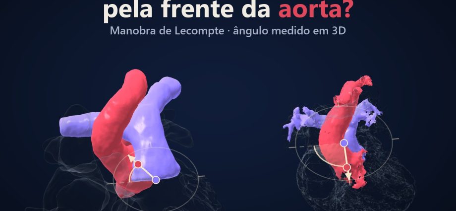
Três corações reais, medidos na mesma régua: no normal, a aorta nasce atrás da pulmonar, a −56°. Numa transposição antes da cirurgia, nasce bem na frente, a +90°. Num terceiro coração, as duas saem lado a lado, a +18°. Qual deles pede a manobra de Lecompte?
📅 Novembro de 2026
Navegue pelo Artigo
Três corações, três ângulos e o que a imagem não consegue mostrar.
- 1. O dilema
- 2. Assista: Por cima da aorta
- 3. Passeio 3D: os três corações
- 4. Os três corações, em graus
- 5. Jatene, 1975. Lecompte, 1981
- 6. Em todos ou só em alguns?
- 7. As coronárias, que a imagem não mostra
- 8. O que vem depois da manobra
- 9. Como foi feito e o que deu errado
- 10. O que é real e o que é esquema
- 11. Perguntas frequentes
- 12. Considerações Finais
- 13. Referências e créditos
1. O dilema
No switch arterial, aorta e pulmonar trocam de lugar. Na transposição mais comum a aorta nasce na frente, e a pulmonar nova ficaria atrás dela. O cirurgião então costuma trazer a pulmonar para a frente da aorta: é a manobra de Lecompte.
E quando as artérias já nascem lado a lado? Aí a resposta deixa de ser óbvia. Uma escola fez a manobra em todos os pacientes; outros grupos escolhem caso a caso. Quis ver essa geometria em corações reais, com um número em vez de um adjetivo.

2. Assista: Por cima da aorta
Um minuto e meio, em diálogo: um ecocardiografista e uma residente. No meio, o filme para e pergunta o que você faria.
3. Passeio 3D: os três corações
Os três corações na mesma escala, lado a lado. Siga os oito passos: o corte axial na altura das raízes, a régua entre os centros da aorta e da pulmonar e a bússola de ângulo mostram −56°, +90° e +18°. No fim, um teste pede que você toque a estrutura certa no coração certo.
Onde nasce a aorta: três corações reais, oito passos.
Normal, transposição antes da cirurgia e artérias lado a lado, na mesma escala. O corte axial na altura das raízes, a régua e a bússola mostram −56°, +90° e +18°. Depois gire, troque de coração e faça o teste.
O modelo só é baixado quando você abre.
4. Os três corações, em graus
Medi o ângulo da aorta em relação à pulmonar no plano axial, na altura das raízes. Zero grau é a aorta ao lado, à direita; +90° é a aorta diretamente à frente; número negativo é a aorta atrás.
| Coração | Origem | Ângulo | Leitura |
|---|---|---|---|
| Normal, 4 anos | HVSMR-2.0, pat5 [1] | −56° | aorta atrás e à direita da pulmonar |
| Transposição antes da cirurgia, idade não informada | NIH 3D Heart Library, modelo #54 | +90° (±5°) | aorta 13 a 15 mm diretamente à frente |
| Artérias lado a lado, 3 anos | HVSMR-2.0, pat40 [1] | +18° | aorta à direita, quase na mesma linha |
Do normal à transposição são 146 graus. No filme, a bússola gira esse caminho ao vivo.
Dois outros corações normais do mesmo banco deram −57° e −63°, o que me deixou mais tranquilo com o −56°. Na transposição, a aorta costuma ficar na frente e à direita, mas a relação varia: lado a lado, ântero-posterior e, às vezes, aorta atrás [2].


5. Jatene, 1975. Lecompte, 1981
A primeira correção anatômica da transposição com sucesso é brasileira. Adib Jatene e colegas, em São Paulo, publicaram o relato preliminar em 1975 [3] e a técnica em 1976 [4]: aorta e pulmonar seccionadas e reanastomosadas, e as coronárias transferidas com um fragmento de parede aórtica.
Seis anos depois, Yves Lecompte e colegas descreveram uma forma de reconstruir a saída pulmonar sem conduto protético [5]. É o que hoje se chama manobra de Lecompte: o tronco pulmonar é deslocado para a frente da aorta [6], e a bifurcação passa a cavalgar a aorta ascendente.
São duas contribuições distintas. Jatene não fez o Lecompte, e Lecompte não inventou o switch.

6. Em todos ou só em alguns?
Aqui a literatura não fecha.
Em 1988, Planché e colegas publicaram 120 neonatos operados com switch: o Lecompte foi feito em todos os 120, inclusive quando os vasos estavam lado a lado [7].
Outros grupos escolhem. Numa série japonesa de 27 pacientes com Taussig-Bing, a manobra foi feita em todos, exceto em 5 com relação lado a lado [8]. Um grupo propôs um ângulo medido na tomografia pré-operatória para escolher a técnica, sobretudo nos casos lado a lado ou oblíquos (14 pacientes, um centro) [9]. Em outra série, o método sem Lecompte apareceu entre os fatores de risco para reintervenção na neopulmonar [10]. Não fazer a manobra também tem custo.
Então, no pat40, a +18°, você faria? Eu não respondo pelo time, e o filme também não.


7. As coronárias, que a imagem não mostra
Boa parte do que decide está fora destes modelos: as coronárias. Nenhum dos três corações tem as coronárias segmentadas.
Num estudo de 255 corações de autópsia, o padrão coronário usual apareceu em 74,8% quando a aorta estava anterior ou ântero-direita, e em 38,9% quando as artérias estavam lado a lado [11]. Numa série de 715 pacientes, as anomalias coronárias (33,7%) se correlacionaram com a relação lado a lado [12].
A posição das artérias, portanto, já avisa um risco. É associação, não regra, e quem decide é o time, com o eco e com o que se vê na sala.

8. O que vem depois da manobra
A estenose pulmonar é a causa mais comum de reintervenção depois do switch [6]. Os autores apontam vários fatores, entre eles o deslocamento anterior do tronco pulmonar no Lecompte, a sutura depois da retirada dos botões coronários e o uso de patch [6]. O Lecompte contribui; não é a única causa.
Os números variam muito de um centro para outro, por isso cada um aqui vale para uma série só:
- em uma série de 688 switches, 11% reintervieram na via de saída do ventrículo direito, e 83% estavam livres dessa reintervenção em 25 anos [13];
- em uma série de 262 neonatos, 10,7% tiveram reintervenção precoce num ramo pulmonar [14].
A relação lado a lado foi fator de risco para reoperação em algumas séries [13, 15]. Em outra, com Taussig-Bing, não houve diferença significativa entre lado a lado e ântero-posterior [16].
9. Como foi feito e o que deu errado
A ideia inicial era usar dois corações do HVSMR-2.0: o pat46 (aorta à frente, +96°) contra o pat40 (lado a lado). Não resistiu à conferência. O pat46 já tem um Glenn, e no nível da raiz a pulmonar dele é um ponto de 42 voxels (cerca de 8 mm³): o +96° media a aorta contra a confluência dos ramos, não valva contra valva. Descartei o caso. Procurei outra transposição limpa, antes do switch, no mesmo banco e não achei (a melhor, pat37, também seguiu com Glenn). Fui buscar o modelo pré-operatório #54 da NIH 3D Heart Library, em domínio público.
O segundo erro foi de método. Os ângulos tinham sido medidos dias antes, sem script salvo. Refiz do zero, agora com script gravado no projeto: os 8 ângulos bateram ao grau.
E o pat40 mudou de papel. Os rótulos do banco mostram átrios invertidos com alça D, o que sugere dupla discordância (transposição congenitamente corrigida). A cirurgia anatômica desse coração seria outra. Ele fica no filme só pela posição das artérias.

10. O que é real e o que é esquema
| Elemento | Origem |
|---|---|
| Corações pat5 e pat40, cortes da ressonância | Real: HVSMR-2.0, segmentação dos autores do banco (lúmen, sem miocárdio) [1] |
| Coração #54 | Malha 3D real (blood pool), NIH 3D Heart Library, sem a imagem-fonte e sem idade informada |
| Ângulos | Calculados: centroides de aorta e pulmonar no plano axial. No #54, ±5° entre cortes, plano do scanner sem correção de inclinação; esquerda e direita são a parte menos garantida |
| Troca das raízes e manobra de Lecompte | Esquema sobre a geometria do #54, marcado como tal. Nenhuma cirurgia mostrada é destes pacientes |
| Coronárias | Não segmentadas; no filme, só esquema tracejado |
| Leitura anatômica | Leitura de IA sobre segmentação e malha; revisão por cardiologista pediátrico humano recomendada |
| Voz | Sintética (Gemini TTS); roteiro e revisão do Dr. Mbula |
Material educativo; as decisões são do médico assistente.
11. Perguntas frequentes
Algum desses corações foi operado com Lecompte?
Não. O pat5 é normal, o #54 é pré-operatório e o pat40 não tem cirurgia registrada no banco.
Lado a lado dispensa o Lecompte?
Não sempre. Há quem tenha feito em todos [7] e quem deixe de fazer em parte dos casos lado a lado [8, 9].
O ângulo serve como critério?
Não. São três corações e um método aproximado. Serve para enxergar a geometria.
A IA fez algum diagnóstico?
Não. Os diagnósticos vêm do banco e da descrição do modelo; os scripts medem posições, e a leitura anatômica é de IA, com os erros contados acima.
Quer ensinar anatomia assim?
A Metodologia AIMED organiza o uso de IA em medicina, com revisão clínica em cada etapa.
12. Considerações Finais
−56°, +90°, +18°. No primeiro coração a pulmonar já passa na frente; no segundo, o cirurgião provavelmente a traria para lá; no terceiro, a pergunta continua aberta, e as coronárias que esta imagem não mostra pesam tanto quanto o ângulo.
Leia também Transposição corrigida em 3D, o episódio sobre a dupla discordância (o que o pat40 provavelmente tem), e Ventrículo único em 3D, o episódio anterior.
Material educativo · as decisões são do médico assistente.
Referências
- Pace DF, Contreras HTM, Romanowicz J, et al. HVSMR-2.0: A 3D cardiovascular MR dataset for whole-heart segmentation in congenital heart disease. Sci Data. 2024;11:721. PMID: 38956063
- Karmegaraj B. Four-dimensional ultrasound imaging of spatial relationship of great arteries in fetuses with TGA. J Ultrasound Med. 2024;43(9):1763-7. PMID: 38752393
- Jatene AD, Fontes VF, Paulista PP, et al. Successful anatomic correction of transposition of the great vessels. A preliminary report. Arq Bras Cardiol. 1975;28(4):461-4. PMID: 1200893
- Jatene AD, Fontes VF, Paulista PP, et al. Anatomic correction of transposition of the great vessels. J Thorac Cardiovasc Surg. 1976;72(3):364-70. PMID: 957754
- Lecompte Y, Zannini L, Hazan E, et al. Anatomic correction of transposition of the great arteries. J Thorac Cardiovasc Surg. 1981;82(4):629-31. PMID: 7278356
- Sobczak-Budlewska K, Łubisz M, Moll M, et al. 30 years’ experience with the arterial switch operation: risk of pulmonary stenosis. Cardiol Young. 2022;33(9):1550-5. PMID: 36040409
- Planché C, Bruniaux J, Lacour-Gayet F, et al. Switch operation for transposition of the great arteries in neonates. A study of 120 patients. J Thorac Cardiovasc Surg. 1988;96(3):354-63. PMID: 3411980
- Masuda M, Kado H, Shiokawa Y, et al. Clinical results of arterial switch operation for double-outlet right ventricle with subpulmonary VSD. Eur J Cardiothorac Surg. 1999;15(3):283-8. PMID: 10333024
- Hongu H, Oda S, Maeda Y, et al. A new predictor of the optimal pulmonary artery reconstruction method during the arterial switch operation. Eur J Cardiothorac Surg. 2023;64(2). PMID: 37140552
- Chen YS, Tsai SK, Chang CI, et al. Prediction of early pulmonary artery stenosis after arterial switch operation: the role of intraoperative transesophageal echocardiography. Cardiology. 2007;109(4):230-6. PMID: 17873486
- Sim EK, van Son JA, Edwards WD, Julsrud PR, Puga FJ. Coronary artery anatomy in complete transposition of the great arteries. Ann Thorac Surg. 1994;57(4):890-4. PMID: 8166537
- Moll M, Michalak KW, Sobczak-Budlewska K, et al. Coronary artery anomalies in patients with transposition of the great arteries and their impact on postoperative outcomes. Ann Thorac Surg. 2017;104(5):1620-8. PMID: 28648541
- Cleuziou J, Vitanova K, Pabst von Ohain J, et al. Incidence and risk factors for right ventricular outflow tract obstruction after the arterial switch operation. Thorac Cardiovasc Surg. 2019;67(1):37-43. PMID: 29715702
- Luo S, Haranal M, Deng MX, et al. Branch pulmonary artery stenosis after arterial switch operation: the effect of preoperative anatomic factors on reintervention. J Thorac Cardiovasc Surg. 2022;164(2):317-327.e8. PMID: 35437174
- Shim MS, Jun TG, Yang JH, et al. Current expectations of the arterial switch operation in a small volume center: a 20-year, single-center experience. J Cardiothorac Surg. 2016;11:34. PMID: 26911711
- Rodefeld MD, Ruzmetov M, Vijay P, et al. Surgical results of arterial switch operation for Taussig-Bing anomaly: is position of the great arteries a risk factor? Ann Thorac Surg. 2007;83(4):1451-7. PMID: 17383356
Dados: HVSMR-2.0 (Pace et al., Sci Data 2024), CC BY 4.0, modificado (pat5 e pat40). Modelo 3D: Case Model #54 d-TGA Pre-Op, Matthew Bramlet, Heart Library, NIH 3D (3DPX-007810), CC0 1.0 (domínio público). Música do filme: ‘Starfire’ by Scott Buckley – released under CC-BY 4.0. www.scottbuckley.com.au. Voz sintética (Gemini TTS: Kore e Zubenelgenubi). Leitura anatômica por IA, revisão humana recomendada.
Why move the pulmonary artery in front of the aorta?
Three real hearts on the same ruler. In the normal heart the aorta arises behind the pulmonary artery, at −56°. In a transposition before surgery it arises right in front, at +90°. In a third heart the two leave side by side, at +18°. Which one calls for the Lecompte manoeuvre?
📅 November 2026
Article Contents
Three hearts, three angles and what the image cannot show.
- 1. The dilemma
- 2. Watch: Over the aorta
- 3. 3D tour: the three hearts
- 4. Three hearts, in degrees
- 5. Jatene, 1975. Lecompte, 1981
- 6. Every case, or only some?
- 7. The coronaries this image can’t show
- 8. What comes after the manoeuvre
- 9. How it was done and what went wrong
- 10. What is real and what is schematic
- 11. FAQ
- 12. Final Thoughts
- 13. References and credits
1. The dilemma
In the arterial switch, the aorta and the pulmonary artery trade places. In the most common transposition the aorta arises in front, so the new pulmonary artery would end up behind it. The surgeon usually brings the pulmonary artery forward, over the aorta: that is the Lecompte manoeuvre.
And when the arteries already arise side by side? Then the answer stops being obvious. One school did the manoeuvre in every patient; other groups decide case by case. I wanted to see that geometry in real hearts, with a number instead of an adjective.

2. Watch: Over the aorta
A minute and a half, as a dialogue between an echocardiographer and a resident. Halfway through, the film stops and asks what you would do.
3. 3D tour: the three hearts
The three hearts on the same scale, side by side. Follow the eight steps: the axial cut at root level, the ruler between the aortic and pulmonary centres and the angle compass show −56°, +90° and +18°. At the end, a test asks you to tap the right structure in the right heart.
Where the aorta arises: three real hearts, eight steps.
Normal, transposition before surgery and side-by-side arteries, on the same scale. The axial cut at root level, the ruler and the compass show −56°, +90° and +18°. Then rotate, switch hearts and take the test.
The model downloads only when you open it.
4. Three hearts, in degrees
I measured the angle of the aorta relative to the pulmonary artery in the axial plane, at root level. Zero means the aorta sits beside it, on the right; +90° means directly in front; a negative number means behind.
| Heart | Source | Angle | Reading |
|---|---|---|---|
| Normal, 4 years | HVSMR-2.0, pat5 [1] | −56° | aorta behind and to the right of the pulmonary artery |
| Transposition before surgery, age not stated | NIH 3D Heart Library, model #54 | +90° (±5°) | aorta 13 to 15 mm directly in front |
| Arteries side by side, 3 years | HVSMR-2.0, pat40 [1] | +18° | aorta on the right, almost level |
From the normal heart to the transposition is 146 degrees. In the film, the compass turns through it live.
Two other normal hearts in the same dataset gave −57° and −63°, which made me more comfortable with the −56°. In transposition the aorta usually sits in front and to the right, but the relationship varies: side by side, anteroposterior and sometimes a posterior aorta [2].


5. Jatene, 1975. Lecompte, 1981
The first successful anatomical repair of transposition came from Brazil. Adib Jatene and colleagues, in São Paulo, published the preliminary report in 1975 [3] and the technique in 1976 [4]: aorta and pulmonary artery divided and reanastomosed, with the coronaries transferred on a fragment of aortic wall.
Six years later, Yves Lecompte and colleagues described a way to rebuild the pulmonary outflow without a prosthetic conduit [5]. That is what we now call the Lecompte manoeuvre: the pulmonary trunk is moved anterior to the aorta [6], and the bifurcation ends up straddling the ascending aorta.
Two separate contributions. Jatene did not do the Lecompte, and Lecompte did not invent the switch.

6. Every case, or only some?
The literature hasn’t settled this.
In 1988, Planché and colleagues reported 120 neonates with an arterial switch: the Lecompte was done in all 120, even when the vessels were side by side [7].
Other groups choose. In a Japanese series of 27 patients with Taussig-Bing anomaly, the manoeuvre was done in all but 5 with a side-by-side relationship [8]. One group proposed an angle measured on preoperative CT to choose the technique, mainly in side-by-side or oblique cases (14 patients, one centre) [9]. In another series, not doing the Lecompte was among the risk factors for reintervention on the neopulmonary artery [10]. Skipping it has a cost too.
So, in pat40, at +18°, would you do it? I don’t answer for the team, and neither does the film.


7. The coronaries this image can’t show
Much of what decides lies outside these models: the coronaries. None of the three hearts has segmented coronaries.
In a study of 255 autopsy hearts, the usual coronary pattern appeared in 74.8% when the aorta was anterior or anterior-right, and in 38.9% when the arteries were side by side [11]. In a series of 715 patients, coronary anomalies (33.7%) correlated with the side-by-side relationship [12].
So the position of the arteries already flags a risk. It’s an association, not a rule, and the team decides, with echo and with what they see in the operating room.

8. What comes after the manoeuvre
Pulmonary stenosis is the most common reason for reintervention after the switch [6]. The authors point to several factors, among them the anterior displacement of the pulmonary trunk in the Lecompte, the suture line after the coronary buttons are harvested and the use of patches [6]. The Lecompte contributes; it isn’t the only cause.
The numbers vary a lot between centres, so each one here holds for one series only:
- in one series of 688 switches, 11% needed reintervention on the right ventricular outflow tract, and 83% were free of it at 25 years [13];
- in one series of 262 neonates, 10.7% had early reintervention on a branch pulmonary artery [14].
A side-by-side relationship was a risk factor for reoperation in some series [13, 15]. In another, in Taussig-Bing, there was no significant difference between side-by-side and anteroposterior [16].
9. How it was done and what went wrong
The first idea was two hearts from HVSMR-2.0: pat46 (aorta in front, +96°) against pat40 (side by side). It didn’t survive the check. Pat46 already has a Glenn, and at root level its pulmonary artery is a 42-voxel speck (about 8 mm³): the +96° measured the aorta against the branch confluence, not valve against valve. I dropped it. I looked for another clean, pre-switch transposition in the same dataset and found none (the best one, pat37, had also gone down the Glenn route). So I went to the public-domain pre-operative model #54 from the NIH 3D Heart Library.
The second mistake was about method. The angles had been measured days earlier with no saved script. I redid them from scratch, this time with the script saved in the project: all 8 angles matched to the degree.
And pat40 changed role. The dataset labels show inverted atria with a D-loop, which suggests double discordance (congenitally corrected transposition). The anatomical operation for that heart would be a different one. It stays in the film for the position of its arteries only.

10. What is real and what is schematic
| Element | Source |
|---|---|
| pat5 and pat40 hearts, MRI slices | Real: HVSMR-2.0, segmentation by the dataset authors (lumen, no myocardium) [1] |
| Heart #54 | Real 3D mesh (blood pool), NIH 3D Heart Library, no source images and no stated age |
| Angles | Calculated: aorta and pulmonary centroids in the axial plane. For #54, ±5° across slices, scanner plane without tilt correction; left and right are the least certain part |
| Root switch and Lecompte manoeuvre | Schematic on the #54 geometry, labelled as such. No surgery shown belongs to these patients |
| Coronaries | Not segmented; shown only as a dashed schematic |
| Anatomical reading | AI reading of segmentations and mesh; review by a human paediatric cardiologist recommended |
| Voice | Synthetic (Gemini TTS); script and review by Dr. Mbula |
Educational material; decisions belong to the treating physician.
11. FAQ
Did any of these hearts have a Lecompte?
No. Pat5 is normal, #54 is pre-operative and pat40 has no surgery recorded in the dataset.
Does side by side rule out the Lecompte?
Not always. Some did it in every case [7]; others skip it in part of the side-by-side cases [8, 9].
Can the angle be used as a criterion?
No. Three hearts and an approximate method. It helps you see the geometry.
Did the AI make a diagnosis?
No. Diagnoses come from the dataset and the model description; the scripts measure positions, and the anatomical reading is AI, with the mistakes told above.
Want to teach anatomy like this?
The AIMED Methodology organises the use of AI in medicine, with clinical review at every step.
12. Final Thoughts
−56°, +90°, +18°. In the first heart the pulmonary artery already runs in front; in the second, the surgeon would probably bring it there; in the third the question stays open, and the coronaries this image can’t show weigh as much as the angle.
Read also Corrected transposition in 3D, the episode on double discordance (what pat40 probably has), and Single ventricle in 3D, the previous episode.
Educational material · decisions belong to the treating physician.
References
- Pace DF, Contreras HTM, Romanowicz J, et al. HVSMR-2.0: A 3D cardiovascular MR dataset for whole-heart segmentation in congenital heart disease. Sci Data. 2024;11:721. PMID: 38956063
- Karmegaraj B. Four-dimensional ultrasound imaging of spatial relationship of great arteries in fetuses with TGA. J Ultrasound Med. 2024;43(9):1763-7. PMID: 38752393
- Jatene AD, Fontes VF, Paulista PP, et al. Successful anatomic correction of transposition of the great vessels. A preliminary report. Arq Bras Cardiol. 1975;28(4):461-4. PMID: 1200893
- Jatene AD, Fontes VF, Paulista PP, et al. Anatomic correction of transposition of the great vessels. J Thorac Cardiovasc Surg. 1976;72(3):364-70. PMID: 957754
- Lecompte Y, Zannini L, Hazan E, et al. Anatomic correction of transposition of the great arteries. J Thorac Cardiovasc Surg. 1981;82(4):629-31. PMID: 7278356
- Sobczak-Budlewska K, Łubisz M, Moll M, et al. 30 years’ experience with the arterial switch operation: risk of pulmonary stenosis. Cardiol Young. 2022;33(9):1550-5. PMID: 36040409
- Planché C, Bruniaux J, Lacour-Gayet F, et al. Switch operation for transposition of the great arteries in neonates. A study of 120 patients. J Thorac Cardiovasc Surg. 1988;96(3):354-63. PMID: 3411980
- Masuda M, Kado H, Shiokawa Y, et al. Clinical results of arterial switch operation for double-outlet right ventricle with subpulmonary VSD. Eur J Cardiothorac Surg. 1999;15(3):283-8. PMID: 10333024
- Hongu H, Oda S, Maeda Y, et al. A new predictor of the optimal pulmonary artery reconstruction method during the arterial switch operation. Eur J Cardiothorac Surg. 2023;64(2). PMID: 37140552
- Chen YS, Tsai SK, Chang CI, et al. Prediction of early pulmonary artery stenosis after arterial switch operation: the role of intraoperative transesophageal echocardiography. Cardiology. 2007;109(4):230-6. PMID: 17873486
- Sim EK, van Son JA, Edwards WD, Julsrud PR, Puga FJ. Coronary artery anatomy in complete transposition of the great arteries. Ann Thorac Surg. 1994;57(4):890-4. PMID: 8166537
- Moll M, Michalak KW, Sobczak-Budlewska K, et al. Coronary artery anomalies in patients with transposition of the great arteries and their impact on postoperative outcomes. Ann Thorac Surg. 2017;104(5):1620-8. PMID: 28648541
- Cleuziou J, Vitanova K, Pabst von Ohain J, et al. Incidence and risk factors for right ventricular outflow tract obstruction after the arterial switch operation. Thorac Cardiovasc Surg. 2019;67(1):37-43. PMID: 29715702
- Luo S, Haranal M, Deng MX, et al. Branch pulmonary artery stenosis after arterial switch operation: the effect of preoperative anatomic factors on reintervention. J Thorac Cardiovasc Surg. 2022;164(2):317-327.e8. PMID: 35437174
- Shim MS, Jun TG, Yang JH, et al. Current expectations of the arterial switch operation in a small volume center: a 20-year, single-center experience. J Cardiothorac Surg. 2016;11:34. PMID: 26911711
- Rodefeld MD, Ruzmetov M, Vijay P, et al. Surgical results of arterial switch operation for Taussig-Bing anomaly: is position of the great arteries a risk factor? Ann Thorac Surg. 2007;83(4):1451-7. PMID: 17383356
Data: HVSMR-2.0 (Pace et al., Sci Data 2024), CC BY 4.0, modified (pat5 and pat40). 3D model: Case Model #54 d-TGA Pre-Op, Matthew Bramlet, Heart Library, NIH 3D (3DPX-007810), CC0 1.0 (public domain). Film music: ‘Starfire’ by Scott Buckley – released under CC-BY 4.0. www.scottbuckley.com.au. Synthetic voice (Gemini TTS: Kore and Zubenelgenubi). AI anatomical reading, human review recommended.
◆ Novidades
- Dor no testículo de madrugada: dá para esperar amanhecer?NOVO
- Criança bateu a cabeça e dormiu: pode deixar dormir? Quando levar ao hospitalNOVO
- Engoliu uma moeda. Aonde ela vai?